Download
1 / 26
310 likes | 647 Views
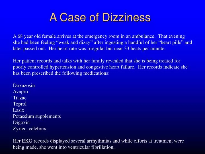
A Case of Dizziness. A 68 year old female arrives at the emergency room in an ambulance. That evening she had been feeling “weak and dizzy” after ingesting a handful of her “heart pills” and later passed out. Her heart rate was irregular but near 33 beats per minute.

E N D
A Case of Dizziness A 68 year old female arrives at the emergency room in an ambulance. That evening she had been feeling “weak and dizzy” after ingesting a handful of her “heart pills” and later passed out. Her heart rate was irregular but near 33 beats per minute. Her patient records and talks with her family revealed that she is being treated for poorly controlled hypertension and congestive heart failure. Her records indicate she has been prescribed the following medications: Doxazosin Avapro Tiazac Toprol Lasix Potassium supplements Digoxin Zyrtec, celebrex Her EKG records displayed several arrhythmias and while efforts at treatment were being made, she went into ventricular fibrillation.
The EKG Physiology in action
Objectives • understand basic cardiac anatomy • understand how cellular action potentials give rise to a signal that can be recorded with extracellular electrodes • understand the path for action potential propagation through the heart • understand the origin of the main phases of electrocardiogram (EKG)
The Heart is a pump has electrical activity (action potentials) generates electrical current that can be measured on the skin surface (the EKG)
At rest, Vm is constant No current flowing Inside of cell is at constant potential Outside of cell is at constant potential A piece of cardiac muscle inside ------------------------------ ++++++++++++++++++ outside - + Currents and Voltages 0 mV
During AP upstroke, Vm is NOT constant Current IS flowing Inside of cell is NOT at constant potential Outside of cell is NOT at constant potential A piece of cardiac muscle AP inside ++++------------------------ ------++++++++++++++ outside current - + Currents and Voltages An action potential propagating toward the positive ECG lead produces a positive signal Some positive potential
inside ++++------------------------ ------++++++++++++++ current - + A negative voltage reading More Currents and Voltages A piece of cardiac muscle An action potential propagating Away from the positive ECG lead produces a negative signal outside
During Repolarization A piece of totally depolarized cardiac muscle A piece of cardiac muscle inside inside +++++++++++++++++++ ------------------------------- ------------+++++++++++ outside Vm not changing No current No ECG signal +++++++------------------- outside current Some negative potential - + More Currents and Voltages Repolarization spreading toward the positive ECG lead produces a negative response
The EKG • Can record a reflection of cardiac electrical activity on the skin- EKG • The magnitude and polarity of the signal depends on • what the heart is doing electrically • depolarizing • repolarizing • whatever • the position and orientation of the recording electrodes
Internodal conducting tissue Cardiac Anatomy Superior venacava Pulmonary veins Atrioventricular (AV) node Left atrium Sinoatrial (SA)A node Atrial muscle Mitral valve Tricuspid valve Purkinje fibers Ventricluar muscle Inferior vena cava Descending aorta
Atrial muscle Internodal conducting fibers Atrial muscle Flow of Cardiac Electrical Activity SA node AV node (slow) Purkinje fiber conducting system Ventricular muscle
Superior venacava Pulmonary veins AV node Left atrium SA node Atrial muscle Mitral valve Specialized conducting tissue Tricuspid valve Purkinje fibers Ventricluar muscle Inferior vena cava Descending aorta Conduction in the Heart approx. 0.44 s 0.12-0.2 s SA node Atria AV node Purkinje Ventricle
approx. 0.44 s 0.12-0.2 s QT PR R T Atrial muscle depolarization P Q S Ventricular muscle repolarization Ventricular muscle depolarization The Normal EKG Right Arm “Lead II” Left Leg
approx. 0.44 s 0.12-0.2 s QT PR Superior ECG venacava Aorticartery SA Pulmonaryartery Pulmonary veins AV node SA node Left atrium Atrial muscle Mitral valve Atria Specialized conducting Interventricular septum tissue Tricuspid valve Purkinje fibers Ventricluar muscle Inferior vena cava Descending aorta Action Potentials in the Heart AV Purkinje Ventricle
A Case of Sudden Death A 68 year old female arrives at the emergency room in an ambulance. That evening she had been feeling “weak and dizzy” after ingesting a handful of her “heart pills” and later passed out. Her heart rate was irregular but near 33 beats per minute. Her patient records and talks with her family revealed that she is being treated for poorly controlled hypertension and congestive heart failure. Her records indicate she has been prescribed the following medications: Doxazosin Avapro Tiazac Toprol Lasix Potassium supplements Digoxin Zyrtec, celebrex Her EKG records displayed several arrhythmias and while efforts at treatment were being made, she went into ventricular fibrillation.
A Case of Sudden Death As noted, the patient’s heart rate was irregular and so were her EKG records. The figures below show two types of patterns seen: