Download
1 / 59
590 likes | 597 Views
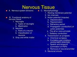
This article explores the structure and functions of nervous tissue in the human body. It covers the organization of the nervous system, the basic cell structure of neurons, and the classification of neurons based on function. Additionally, it discusses the structure of nerves and provides an overview of the spinal cord and its associated structures.

E N D
Function of the Nervous System • Controls and integrates all body activities within limits that maintain life (homeostasis) • Three basic functions • Sensing changes with sensory receptors • Fullness of stomach or sun on your face • Interpreting and remembering those changes • Reacting to those changes with effectors • Muscular contractions • Glandular secretions Afferent – Sensory (acending) Efferent – Motor (decending)
Organization of the Nervous System • Two major anatomical subdivisions • Central nervous system (CNS) • Brain and spinal cord enclosed in bony coverings • Peripheral nervous system (PNS) • Nerve • Bundle of nerve fibers in connective tissue • Ganglion • Swelling of cell bodies in a nerve
Basic Cell Structure of a Multipolar Neuron Dendrites: Branching extensions of cell body providing structural pathway for afferent information Nucleus: DNA-containing structure of cell body Axon: structural pathway for efferent information Axon hillock Cell body: constitutes nucleus and cytoplasm Axon hillock: conic area of origin of axon from nerve cell body
Basic Cell Structure of a Multipolar Neuron Axon terminals(synaptic end bulbs): axon terminates, communicates with subsequent cells Node of Ranvier: gap in myelin sheath allow formation of new AP, increases rate of impulse transmission Myelin: insulates and rapid transmission of impulses Telodendria Telodendria: distal branching of neuronal axon Schwann Cell Nissl bodies Schwann cell: cell that surround and myelinate axons in PNS Synapse: junction between axon terminals of neurons or the effector organ where neural impulses are transmitted Nissl bodies: RER of neurons, site of protein synthesis
Anatomy of Multipolar Neuron Review Axon terminals/synaptic end bulbs 10. Axon hillock 3. Dendrites 4. 9. Telodendria Cell body (neurosoma) 1. Schwann Cell 6. Node of Ranvier (neurofibril node) 8. 5. Axon 11. Nissl bodies Myelin sheath (neurolemma) 7. 2. Nucleus
Neurons Classified by Function • Sensory (afferent) neurons • Sensory receptors detect changes in body and external environment • Transmit impulses to brain or spinal cord • Interneuron (association) neuron • Connect motor and sensory neurons in neural pathways (CNS) • 90% of our neurons are interneurons • Process, store and retrieve information • Motor (efferent) neuron • Carries impulses away from CNS to viscera, muscles and glands
Structure of a Nerve • Spinal nerve • 1 of 31 pairs of nerves originating from spinal cord • Epineurium • Covering of entire nerve • Perineurium • Surrounds group of nerve fibers forming a fascicle • Endoneurium • Wrapping of each nerve fibers • Fascicle • Small bundle of nerve fibers • Axon • Long and single nerve-cell process – nerve fiber • Myelin sheath • Fatty white substance surrounding axon (nerve fiber)
Nervous Tissue Review • Using the nerve models, identify the basic cell structures of the multipolar neuron listed in 1.1 of Survival Guide. • Observe the following slides under the microscope and be able to identify: • Multipolar neuron • Structures of a peripheral nerve (see items a-f of 1.6)
Terminology and Anatomy of the Spinal Cord • Tracts - collections of axons in CNS • Nerves - collections of axons in PNS • Ganglia - collections of neuron cell bodies in PNS • Nucleus (pl. nuclei) - collections of neuron cell bodies in CNS
Structures of the Spinal Cord • Ventral root (anterior root) • Efferent = motor • Dorsal root (posterior root) • Afferent = sensory • Dorsal root ganglion (posterior root ganglion) • Swelling lateral of SC containing sensory neuron cell bodies • Spinal nerve • Merging of ventral and dorsal roots (mixed nerve)
Structures of the Spinal Cord • ~ 2 cm in diameter • Extends through vertebral canal to the L1 or L2 vertebral level • Conus medullaris (L1-2) • Conical, tapered, and distal end of spinal cord • Cauda equina (horse’s tail) • Bundle of spinal nerves and nerve roots • Filum terminale (terminal thread) • Longitudinal support to spinal cord
Gray and White Matter Divisions of Spinal Cord • 3 gray matter horns • Posterior gray horns • Contain somatic and visceral sensory nuclei. • Anterior gray horns • Contain somatic motor nuclei. • Lateral gray horns • Contain visceral motor nuclei (only located in the thoracic and lumbar segments). • 3 white matter columns • Posterior white column • Anterior white column • Lateral white column • Each column contains tracts (axons) • Ascending tracts carry sensory information from the body toward the brain. • Descending tracts carry motor commands to the spinal cord.
Spinal Cord: Gray Matter Central Canal (through which CSF flows) Posterior Gray Horn Anterior and Posterior gray commissures Lateral Gray Horn Anterior Gray Horn The central “butterfly” or gray matter contains cell bodies, neuroglia, dendrites and unmyelinated axons (interneurons).
Spinal Cord: White Matter Posterior white column Posterior median sulcus Lateral white column Anterior white commissure Anterior median fissure Anterior white column White columns contain tracts (fasciculus) relaying same type information in same direction: ascending tracts carry sensory information and motor information by the descending.
Spinal Cord: Building the Nerve • DR= sensory neurons to SC • VR = motor neurons to somatic and visceral effector • DRG = cell bodies of sensory neurons Dorsal (posterior) root Dorsal root ganglion Dura and Arachnoid matter Ventral (anterior) root Pia matter Spinal nerve
Disease of the Spinal Cord: Poliomyelitis • Polio =gray matter (of the brain or spinal cord) • Viral infection • Inflammation of gray matter in anterior gray horn motor neurons • Neurons innervate muscles • Signs and symptoms • Initial flu like symptoms • Muscle ache and weakness • Ultimately muscle paralysis
Disease of the Spinal Cord:Amyotrophic Lateral Sclerosis • Amyotrophic = without muscle nourishment • ALS (AKA: Lou Gehrig’s Disease) • Progressive neurodegenerative disease. • Affects nerve cells in brain and spinal cord specifically anterior gray horn motor neurons and precentral gyrus. • Idiopathic, but thought to be genetic. • Signs and symptoms • Loss of ability to initiate and control muscle movement. • Voluntary muscle action progressively affected leading to loss in ability to speak, eat, move and breathe. • Leads to paralysis and death.
Spinal Cord Review • Using the spinal cord models, identify the structures listed in Spinal Cord #9 of Survival Guide. • Observe the following slides under the microscope and be able to identify: • Structures listed in Spinal Cord #9 of Survival Guide.
31 Pairs of Spinal Nerves • 8 cervical spinal nerves (C1-C8) • C1-C7 emergeabove vertebra for which named • C8 emergesbetween C7 and T1 • 12 thoracic spinal nerves (T1-T12) • Emerge below vertebra for which named • 5 lumbar spinal nerves (L1-L5) • Emerge below vertebra for which named • 5 sacral spinal nerves (S1-S5) • Emerge below vertebra for which named • 1 coccygeal pair (coccyxgeal) • Emerge below vertebra for which named
From Spinal Roots to Nerve Plexuses • Dorsal and ventral roots exit the spinal cord and join together to make a spinal nerve • The spinal nerve splits into dorsal ramus and ventral ramus • Some ventral rami give off branches to sympathetic ganglion • Other ventral rami mix and match to make up nerve plexuses except for thoracic
Spinal Cord Ventral Roots (motor) Dorsal Roots (sensory) Spinal Nerve(mix sensory and motor) Communicating Rami(white ramus and gray ramus) Dorsal Ramus(mixed) Ventral Ramus(mixed) Sensory/Motor Back Skin & Muscles Nerve Plexuses Sympathetic ganglia
Ventral Rami Form Plexuses • Dorsal rootonly contains sensory neurons going toward the spinal cord. • Ventral rootonly contains motor neurons going out of the spinal cord. • Ventral ramicontain both sensory and motor neurons. • Plexus • Branching network of intersecting nerves • Roots, trunks, divisions, cords, branches • Cervical • Brachial • Lumbar • Sacral • Coccygeal
Cervical Plexus: formed by C1-C4 Phrenic nerve is an important branch of cervical plexus supplying the diaphragm. “C3, 4, 5 keeps the diaphragm alive”
1. Musculocutaneous nerve (C5-T1) 2. Axillary nerve (C5-C6) 3. Radial nerve (C5-T1) 4. Median nerve (C6-T1) 5. Ulnar nerve (C8-T1) Brachial Plexus: formed by C5-C8 and T1
Nerves of the Brachial Plexus • Axillary nerve (C5-C6) • Motor: deltoid and teres minor muscles • Sensory: skin of the shoulder • Musculocutaneous nerve (C5-T1) • Motor: flexor muscles of the arm • Sensory: lateral surface of the forearm
Nerves of the Brachial Plexus • Radial nerve (C5-T1) • Motor: muscles of posterior arm and forearm • Sensory: posterolateral hand including 1st digit • Median nerve (C6-T1) • Motor: flexor muscles on radial side of forearm • Sensory: anterolateral surface of hand (thenar) and posterior 2nd and 3rd , and ½ of 4th digits. • Ulnar nerve (C8-T1) • Motor: flexor muscles of forearm and hand on ulnar side • Sensory: anteromedial (hypothenar) and posteromedial surface of the hand, ½ of 4th and 5th digits.
Lumbar Plexus: formed by L1-L4 and contributions from T12 Major nerves: 1. Femoral nerve (L2-L4) and2. Obturator nerve (L2-L4)
Major Nerves of the Lumbar Plexus • Femoral nerve (L2-L4 ) • Motor: quadriceps group, pectineus and iliopsoas mm • Sensory: anterior-medial thigh and medial surface of leg and foot • Obturator nerve (L2-L4) • Motor: adductors of hip • Sensory: medial surface of thigh
Sacral Plexus: formed by L4 - L5 and S1-S4 Innervates buttock, lower limb, pelvis, and perineum.
Major Nerves of the Sacral Plexus • Sciatic nerve (L4–S3) • Common fibular and tibial nerves collectively known as sciatic nerve. • Branches behind knee: • Common fibular nerve • Motor: lateral and anterior muscles of the leg • Tibial nerve • Motor: posterior muscles of the leg Common Fibular Nerve
Major Nerves of the Sacral Plexus • Pudendal nerve (S2-S4) • Motor: muscles of the perineum (pelvic floor mm.) • Sensory: perineum, genitalia
Coccygeal Plexus: formed by S4, S5, and Co1 • Ventral branches of the Co1 and S5 nerves • Communication from S4 • Give rise to anococcygeal nerves
Sympathetic Chain Ganglia • Part of the sympathetic nervous system. • Nervous mechanism, which enables communication alerting body to stress and impending danger (fight-or-flight). • Found extensively in thoracic and lumbar regions. • Ventral root gives rise to a myelinated preganglionic fiber (white rami) to sympathetic chain ganglia • Fibers may synapse white rami, in collateral ganglia, or adrenal medulla.
Scheme showing structure of a typical spinal nerve. 1. Somatic efferent. 2. Somatic afferent.3,4,5. Sympathetic efferent. 6,7. Sympathetic afferent.
Nerve, and Nerve Plexus Review • Using the nerve and extremity muscle models, identify the structures listed in Spinal Cord images 1.8-1.14 of Survival Guide.
Reflexes: Terminology and Definitions • Regional affects of reflexes • Ipsilateral • Affecting same side of body as stimulus. • Contralateral • Affecting opposite side of body from stimulus. • Consensual • Affects both sides of body simultaneously. • Development of reflexes • Innate reflex • Connection that form between neurons during development (sucking, grasping, removing hand from hot stove). • Acquired reflex • Complexed learned motor patterns enhanced by repetition (foot on brake when trouble appears).
Reflexes: Terminology and Definitions • Nature of response • Visceral (autonomic) reflex • Controls actions of smooth muscle, glands, organs, and adipose tissue (digestion, blood pressure). • Somatic reflex • Control skeletal muscle contractions (include superficial stretch reflexes). • Complexity of circuit • Monosynaptic reflex • Simplest reflex arc. • Sensory neuron synapses directly on a motor neuron. • Polysynaptic reflex • Complexed with one or more interneuron between sensory and motor neurons • Interneurons involved can control several muscle groups and produce either excitatory or inhibitory polysynaptic potentials
Reflexes: Terminology and Definitions • Processing site • Spinal reflexes • Important interconnections and processing events occur in spinal cord. • Cranial reflexes • Integration center located in brain. • Motor neurons • Upper motor neurons (UMN) – “uninhibited” • Motor cortex of brain and terminate within the medulla or spinal cord. • Damage: results in spasticity and exaggerated reflexes: “spastic paralysis”. • Lower motor neurons (LMN) – “disconnected” • Cell bodies in spinal cord and terminate in skeletal muscle. • Damage: weakness, fasciculation and atrophy: “flaccid paralysis”.
Reflexes: Rapid, Automatic Responses to a Stimulus • Critical role in protecting body in the regulation of homeostasis. • Reflex arc • Neuronal pathway of a reflex • 5 necessary components • Receptor • Sensory neuron • Integration center • Motor neuron • Effector
Reflex Testing: Evaluating Nervous System’s Integrity • Intact sensory afferent nerves to spinal cord. • Intact synapse within spinal cord. • Intact efferent motor nerves from spinal cord. • Adequately functioning muscle. • Reflex testing is important in the clinical evaluation of nervous system integrity. • Abnormality in a reflex indicates dysfunction within reflex arc. • Hyporeflexic • Less than average response. • May be indicative of a metabolic disease - hypothyroidism. • Hyperreflexic • Exaggerated response. • May be a sign of CNS damage.
Spinal Reflexes: Stretch Reflex (myotatic) • Stretching of muscle activates a muscle spindle. • Bundle of specialized skeletal muscle fibers that act as sensory receptors. • Impulse transmitted by afferent fibers to spinal cord. • Motor neurons in spinal cord cause stretched muscle to contract. • Integration center causes antagonist muscle to relax (reciprocal inhibition). • Monosynaptic reflex • Interpretation: • Decreased reflexes should lead to suspicion that reflex arc has been affected. • Possible sensory nerve fiber or spinal cord gray matter (motor fiber), LMN lesions.
Patellar Reflex: L2, L3, L4 (knee jerk) • Subject sits on bench or chair comfortably with knee bent and foot off floor. • Sharply strike patellar tendon between inferior aspect of patella and insertion. • Leg extends • Interpretation • Demonstrates sensory and motor connections between muscle and spinal cord are intact.
Ankle Reflex: S1, S2 (ankle jerk) • Subject sits on bench or chair comfortably with knee bent and foot off floor. • Stretch Achilles tendon pushing up slightly with hand on ball of foot (dorsiflex ankle). • Sharply strike reflex hammer onto tendon above calcaneus. • Measure response by feeling downward push against hand (plantar flexion). • Interpretation • Demonstrates sensory and motor connections between muscle and spinal cord are intact.
Biceps Reflex: C5, C6 (biceps jerk) • Bend subject’s arm elbow so it is relaxed and supported. • Locate tendon of biceps (antecubital fossa). • Traction tendon by pulling inferiorly with thumb. • With opposite hand, strike thumb sharply with reflex hammer. • Interpretation • Demonstrates sensory and motor connections between muscle and spinal cord are intact.
Spinal Reflexes: Golgi Tendon Reflexes • Opposite of stretch reflex. • Monitors external tension produced during muscular contraction (compare to stretch reflex muscle spindle is length) • Prevents tearing or breaking of tendons. • Increase in muscle tension activates receptor in tendon (Golgi tendon organ). • Muscle relaxes and lengthens in response to its antagonist's contraction. • Deep Tendon (DT) reflex inhibits agonist and excites antagonist.