Download
1 / 48
480 likes | 599 Views
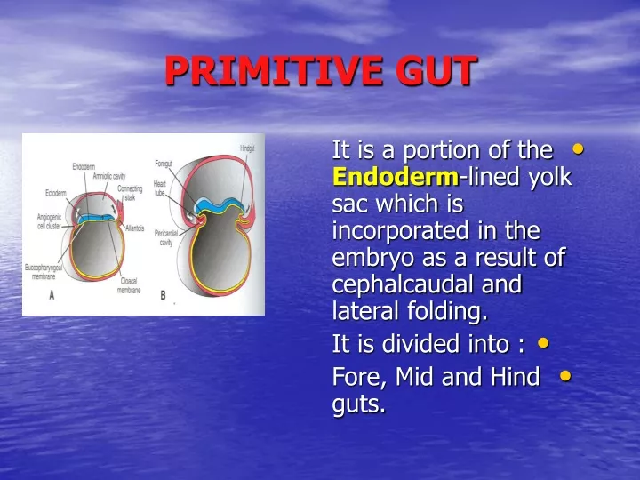
PRIMITIVE GUT. It is a portion of the Endoderm -lined yolk sac which is incorporated in the embryo as a result of cephalcaudal and lateral folding. It is divided into : Fore, Mid and Hind guts. PRIMORDIAL GUT.

E N D
PRIMITIVE GUT • It is a portion of the Endoderm-lined yolk sac which is incorporated in the embryo as a result of cephalcaudal and lateral folding. • It is divided into : • Fore, Mid and Hind guts.
PRIMORDIAL GUT • It gives rise to most of the Epithelial lining and Glands of the digestive tract. • The muscular and connective tissue is derived fro the Splanchnicmesoderm. • The epithelium at the cranial end (Stomodeum) and the caudal end (Proctodeum) is derived from Ectoderm.
FORE GUT • It is the part incorporated in the cephalic part of the embryo. • It forms a blind tube which lies caudal to the pharyngeal tube. • It extends as far caudally as the primordium of the liver.
DERIVATIVES OF THE FORE GUT • 1. Primordial pharynx (oral cavity, tongue, salivary glands, tonsils and upper respiratory system). • 2. Lower respiratory system. • 3. Esophagus and Stomach. • 4. Liver and biliary apparatus.
ESOPHAGUS • Origin: • It develops caudal to the primitive pharynx. • The Tracheoesophageal septum divides the fore gut into a Ventral portion (RespiratoryDiverticulum) and a Dorsal portion (Esophagus).
LENGTH • Initially the esophagus is short. • It elongates because of the growth and descent of the heart and lungs. • The final length is reached by the seventh week.
STRUCTURE • The epithelial lining and glands are from the Endoderm. • Muscles : • Upper third : Striated. From the mesenchyme • in the caudal pharyngeal arches. • Lower third : Smooth. • From splanchnic mesoderm.
ESOPHAGUS • Rapid proliferation of the epithelium obliterates the lumen temporarily. • Recanalization of the esophagus normally occurs at the end of the embryonic period.
STOMACH • It appears in the middle of the (4TH ) week as a fusiform dilatatation at the caudal end of the foregut. • It is initially in the median plane.
SHAPE • Its Cranial and Caudal ends are in the median plane. • It has Dorsaland a Ventral borders. • The dorsal border grows faster than the ventral. • This demarcates the Greater Curvature of the stomach.
ROTATION • The stomach rotates (90) degrees in a Clockwise direction around its longitudinal axis.
RESULTS OF ROTATION • (1) Lesser Curvature moves to the Right. • The Greater Curvature moves to the Left. • (2) The Left side becomes the Ventral surface. • The Right side becomes the Dorsal surface.
RESULTS OF ROTATION • This explains the Left Vagus nerve supplies the anterior wall of the adult stomach and the RightVagus supplies the posterior wall.
RESULTS OF ROTATION • (3) The Cranial region moves to the Left and Inferiorly. • The Caudal region (pyloric) end moves to the Right and Superiorly.
RESULTS OF ROTATION • (4) The Long axis of the stomach becomes almost Transverse to the Long axis of the body.
MESENTRIES • The stomach is attached to the dorsal body wall by the Dorsal Mesogastrium and to the ventral body wall by the VentralMesogastrium.
OMENTAL BURSA • Isolated intercellular clefts appear in the Dorsalmesogastrium. • These clefts fuse and form a single cavity (Lesser Peritoneal Sac). • Rotation of the stomach pulls the Dorsal mesogastrium to the left and enlarges the bursa.
OMENTAL BURSA • The bursa represents the extension of the right side of the peritoneal cavity behind the stomach. • The developing diaphragm cuts off the superior part of the bursa. If it persists, it lies medial to the base of the right lung.
OMENTAL BURSA • The persisting bursa forms a closed sac (InfraCardiac bursa). • Superior Recess of the omental bursa: • It represents the inferior portion of the superior part of the bursa.
OMENTAL BURSA • Inferior Recess of omental bursa: • It is developed due to the enlargement of the stomach. • It is found between the layers of the elongated part of the dorsal mesogastrium (GreaterOmentum).
OMENTAL BURSA • The omental bursa communicates with the peritoneal cavity through the OmentalForamen.
DUODENUM • It develops from the caudal par of the Foregut and the cranial part of the Midgut. • The junction of the two guts is caudal to the origin of the Bile Duct.
DUODENUM • The duodenum forms a C- shaped loop. • By the Rotation of the stomach, the duodenal loop moves to the Right. • This rotation and the rapid growth of the head of the pancreas swings the duodenum from the initial midline position to the left side of the abdominal cavity.
RETROPERITONEAL POSITION • Initially the duodenum has a Dorsal mesoduodenum. • It fuses with the peritoneum of the posterior abdominal wall. • Later the two layers disappear and the duodenum becomes fixed.
RECANALIZATION • In the 4th and 5th weeks, the lumen is temporarily obliterated by Proliferation of the epithelial cells. • Degeneration of the cells causes normal vacuolation and the duodenum becomes recanalized.
SPLEEN • It is a large vascular lymphatic organ. • It is derived from a mass of Mesenchymalcells between the layers of the Dorsal Mesogastrium. • It is developed during the (5th ) week. • It acquires its characteristic shape in the fetal period.
SPLEEN • The Fetal spleen is Lobulated. • The lobules disappear Beforebirth. • The notches in the upper border of the adult spleen are remnants of the grooves that separated the fetal lobules.
SPLEEN • Because of the Rotation of the stomach, the portion between the spleen and the dorsal midline swings to the left. • It fuses with the peritoneum over the left kidney.
SPLEEN • The peritoneum along this line of fusion degenerate. • The spleen is connected to the body wall in the region of the left kidney by the lienrenal ligament. • This explains the tortuous course of the adult splenic artery.
HEPATIC DIVERTICULUM • It arises as a ventral outgrowth from the caudal part of the fore gut during the (4th ) week. • It extends into the septum transversum. • Its larger cranial part is the primordium of the Liver. • The small caudal part becomes the GallBladder.
SEPTUM TRANSVERSUM • It is a mass of Splanchnic mesoderm between the developing heart and the mid gut.
DERIVATIVES OF SEPTUM TRANSVERSUM • 1. Central tendon of the diaphragm. • 2. Ventral mesentery with its two parts : • (a) Falciform ligament. • (b) Lesser omentum. • 3. Kupffer cells and fibrous tissue of the liver. • 4. Visceral peritoneum of the liver.
STRUCTURE OF THE LIVER • The proliferating Endodermal cells give rise to : • 1. Parenchyma (liver cells). • 2. Epithelial lining of the (Intrahepatic) portion of the biliary apparatus.
STRUCTURE OF THE LIVER • The Vitelline and Umblical veins will form • the Hepatic Sinusoids. • The development and functional segmentation of the liver is determined by the quantity of oxygenated blood passing to the liver through the umbilical vein.
SIZE • The liver fills a large part of the abdominal cavity between (5th -10th ) weeks. • It accounts about (10%) of the total weight of the fetus by the 9th week. • It is formed of right and left lobes of almost the same size. • The right lobe soon becomes the larger one.
FUNCTION • Hematopoiesis begins during the 6th week. • It is responsible for : • 1. Bright reddish appearance of the liver. • 2. Large size of the liver (7th -9th ) weeks. • Bile formation begins during (12th ) week.
EXTRA HEPATIC BILIARY APPARATUS • Ahepatic ductcomes out from each lobe. • The two ducts unite to form asingleCommonHepatic duct. • Bile duct: • The connection between the hepatic diverticulum and the fore gut (duodenum) narrows to form the bile duct. • Gall bladder and Cystic duct: • Formed from a small ventral outgrowth of the bile duct.
EXTRA HEPATIC BILIARY APPARATUS • The bile duct initially opens into the Ventral aspect of the duodenal loop. • Growth and Rotation of the duodenum brings this opening Dorsally.
EXTRA HEPATIC BILIARY APPARATUS • The proliferating epithelial cells cause temporary occlusion of the biliary apparatus. • Recanalization is restored by degeneration of these cells. • Bile enters the duodenum during (13th ) week. It gives the meconium its dark green color.
PANCREAS • It is formed from two Pancreatic Buds (Dorsal and Ventral). • They arise from Endodermal cells in the proximal part of the duodenum (caudal part of the foregut) between the layers of the dorsal and ventral mesentery respectively.
PANCREATIC BUDS • The Dorsalbud is the larger. • It appears first and is slightly cranial to the ventral bud. • The Ventral bud develops near the entry of the bile duct into the duodenum.
ROTATION • The Rotation of the duodenum to the right and its C shaped appearance shifts the entry of the bile duct and the ventral pancreatic duct Dorsally.
FUSION • The ventral bud comes to lie immediately below and dorsal to the dorsal bud. • The parenchyma of the buds fuse and their ducts anastomose.
DERIVATIVES OF THE BUDS • The Ventral bud forms : • 1.The Uncinate process. • 2. The Inferior part of the head. • The Dorsal bud forms the remaining part of the pancreas.
DUCTS • Main pancreatic duct • It is formed from the distal part of the dorsal pancreatic duct and the entire ventral pancreatic duct. • It opens with the bile duct into the Major duodenal papilla.
DUCTS • Minor pancreatic duct : • It is the Proximal part of the dorsal pancreatic duct (if persists). • It opens into the Minorduodenal papilla (2 cm) cranial to the main duct.
FUNCTIONS • (1) Insulin secretion • Pancreatic islets ( of Langerhans) develop from the parenchymatous pancreatic tissue. • They produce insulin by the 10th week.
FUNCTIONS • (2) Glucagon and Somatostatin. • Their containing cells develop before differentiation of the insulin secreting cells. • Glucagon has been detected in the fetal plasma at 15 weeks.