Download
1 / 58
580 likes | 599 Views
Explore the concepts of gene mutations at the molecular and chromosomal levels, their effects on protein structure, and how they can lead to diseases like sickle cell anemia, thalassemia, Ehlers-Danlos syndrome, and Alzheimer's disease. Learn about mutation rates, mutagenesis, induced mutations, and examples of mutagens. Discover how mutations are crucial for evolution and why understanding them is essential for disease treatment.

E N D
Human Genetics Concepts and Applications Eighth Edition Powerpoint Lecture Outline Ricki Lewis Prepared by Dubear Kroening University of Wisconsin-Fox Valley
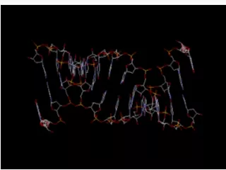
Mutation • A change in the nucleotide sequence of a gene (< 1%), polymorphism (>1%) • May occur at the molecular or chromosomal level • The effect of mutations vary • Mutant refers to an unusual phenotype • Mutations are important to evolution
Somatic Mutations • Occur in cells of the body, excluding the germline • Affects subsequent somatic cell descendants • Not transmitted to offspring Germline Mutations • Mutations that occur in the germline cells • Possibility of transmission to offspring
Mutations Alter Proteins • Examples of mutations that cause disease: • Beta globin gene • Collagen • Early onset Alzheimer
Hemoglobin • Four globular proteins surrounding heme group with iron atom: two beta chains and two alpha chains • Function is to carry oxygen in red blood cells from lungs to body and carbon dioxide from cells to lungs
Mutations Can Cause Cells to Sickle Chapter 12 Opener
Sickle Cell Disease Results from a Base Change in Beta Globin Gene Figure 12.2
Sickle Cell Anemia • Mutation encodes valine in place of glutamic acid • Phenotype associated with homozygotes • Altered surface of hemoglobin allows molecules to link in low oxygen conditions • Creates sickle shape of RBC • Sickling causes anemia, joint pain, and organ damage when RBC become lodged in small blood vessels
Thalessemia • Caused by another beta hemoglobin mutation • Too few beta hemoglobin molecules • Excess of alpha hemoglobin leads to iron release, which destroys RBC, damages heart, liver, and endocrine glands. • Thalassemia minor (heterozygous) • Thalassemia major (homozygous for mutation and more severe)
Collagen Comprises • 60% of protein in bone and cartilage • A significant proportion of skin, ligament, tendon, tooth dentin, and connective tissue Has a precise structure • Triple helix of two alpha1 and one alpha 2 proteins • Longer precursor, procollagen is trimmed to form collagen
Ehlers-Danlos Syndrome Figure 12.4 A mutation prevents procollagen chains from being cut
Alzheimer Disease • Mutations in presenilin 1 cause early-onset autosomal dominant Alzheimer disease • Found on chromosome 14 • Presenilin protein is a receptor anchored in the Golgi membrane • Monitors beta amyloid usage • Missense mutations in presenilin result in beta amyloid accumulation.
One Cause of Alzheimer Disease Figure 12.5
How Mutations Cause Disease • Understanding the molecular cause of a disease may assist in treatment • The same symptoms may be caused by different mutations • Table 12.2 lists several examples of mutations and the diseases they produce
Mutations • Change in the DNA • May occur spontaneously or by exposure to a radiation or chemicals • An agent that causes a mutation is a mutagen
Spontaneous Mutation • De novo or new mutations • Not caused by exposure to known mutagen • Errors in DNA replication • DNA bases have slight chemical instability • Exist in alternating forms called tautomers • As replication fork encounters unstable tautomers, mispairing can occur
Spontaneous Mutation Figure 12.6
Spontaneous Mutation Rate • Rate differs for different genes • Vary by size • Sequence dependence • Hot spots • Table 12.3 lists rates for several genes • On average, 1/100,000 each round of replication • Each individual has multiple new mutations • Most by are not in coding regions of genes
Determining Mutation Rate • Estimates of spontaneous mutation rate can be derived from observation of dominant traits. • For autosomal genes, mutation rate = # of cases 2 (# of individuals)
Mutational Hot Spots • Short repetitive sequences • Pairing of repeats may interfere with replication or repair enzymes • Palindromes • Often associated with insertions or deletions • Duplications of larger regions • Mispairing during meiosis
DNA Symmetry May Increase Mutation Rate Figure 12.7
Gene Duplication May Increase Mispairing Figure 12.8
Induced Mutations • Caused by mutagens, many are also carcinogens and cause cancer Examples: • Alkylating agents: remove a base • Acridine dyes: add or remove base • Xrays: break chromosomes delete a few nucleotides • UV radiation: creates thymidine dimers • Site-directed mutagenesis:
Ames Test • An in vitro test of the mutagenicity of a substance • One version uses Salmonella bacteria with mutation in gene for histidine • Bacteria are exposed to test substance • Growth on media without histidine is recorded • Bacteria only grow if mutations have occurred • Substance can be mixed with mammalian liver tissue prior to testing to mimic toxin processing in humans
Exposure to Mutagens • Workplace • Industrial accidents • Chernobyl • Medical treatments • Weapons • Natural sources
Sources of Radiation Exposure Table 12.5
Point Mutations A change of a single nucleotide • Transition purine replaces purine A to G or G to A or pyrimidine replaces pyrimidine C to T or T to C • Transversion purine replaces pyrimidine or pyrimidine replaces purine A or G to T or C T or C to A or G
Missense Mutation • A point mutation that changes the codon • Causes a substitution of an amino acid • Missense mutations may affect protein function severely, mildly, or not at all. Example: • Hemoglobin mutation • Glutamic acid to valine causes sickle cell anemia
Nonsense Mutation • A point mutation changing a codon for an amino acid into a stop codon • Creates truncated proteins that are often nonfunctional • Some have dominant effects due to interference with normal functions Example: • A factor XI deficiency is a nonsense mutation changing glutamic acid to a “stop” • Short protein cannot function in clotting
Splice Site Mutations • Alters a site where introns are normally removed • Intron translated or exon skipped • Examples • CF mutation • BRCA1 • Familial dysautonomia (FD)
Insertions or Deletions • The genetic code is read in triplet nucleotides • Addition or subtraction of nucleotides not in multiples of three leads to a change in the reading frame • Causes a frameshift and alters amino acids after mutation • Addition or subtraction of nucleotides in multiples of three leads to addition or subtraction of entire amino acids
Examples • 2/3 of Duchenne muscular dystrophy cases are large deletions • Gaucher disease is caused by a single base insertion creating a frameshift • A tandem duplication is an insertion of identical sequences side by side • Charcot-Marie-Tooth disease is caused by a tandem duplication of 1.5 million bases
Pseudogenes • A DNA sequence similar to a gene but which is not translated • May or may not be transcribed • May have evolved from original gene by duplication and acquired mutation • Crossing over between a pseudogene and functional gene can disrupt gene expression
Expanding Repeats • Insertion of triplet repeats leads to extra amino acids • Some genes are particularly prone to expansion of repeats • Number of repeats correlates with earlier onset and more severe phenotype • Anticipation is the expansion of the triplet repeat with an increase in severity of phenotype with subsequent generations
Myotonic Dystrophy: A Triplet Repeat Disease Figure 12.10
Triplet Repeat Disorders Table 12.7
Copy Number Variants • Sequences in more than one place in a genome • Range from a few bases to millions • Affects susceptibility to HIV • Comparative genomic hybridization
Importance of Position The degree that a mutation alters phenotype depends on • Where in the gene the change occurs • How it affects conformation or expression • Examples – hemoglobin and prions
Globin Mutations Table 12.8
Prion Disorders • Caused by mutation in the prion gene • Leads to an abnormally shaped prion protein • The mutant form converts normal prion proteins to mutant protein shapes • Can be inherited or transmitted like an infection from tissue with the mutant protein • Example • In cows a mutant prion protein causes bovine spongiform encephalopathy (“mad cow”) • Humans develop Creutzfeldt-Jakob disease by eating beef with mutant prions
Developing Prion Diseases • Amino acid in 129th position is key to developing disease • Individuals homozygous with valine (VV) or methionine (MM) develop disease • Heterozygotes have normal function • Position 178 is also important due to the folding of the protein
Not All Mutations Impact Protein Function • Silent mutationsare mutations that do not alter the amino acid encoded Example: • A mutation from AAA to AAG alters the DNA, the protein sequence remains unchanged • AAA and AAG both encode for lysine • These codons are called synonymous codons
Not All Mutations Impact Protein Function • Missense mutations alter the encoded amino acid to another amino acid • Creates a nonsynonymous codon • Some nonsynonymous mutations are conservative; chemically similar amino acid and may not alter function • The impact of a missense mutation is not predictable from protein sequence alone
Not All Mutations Impact Protein Function • Conditional mutationsproduce a phenotype under particular conditions or environments • G6PD enzyme responds to oxidants, chemicals that strip electrons from other molecules • High levels of oxidants occur when eating fava beans or taking antimalarial drugs Conditions Individuals with mutations in G6PD Low oxidants no phenotype High oxidants red blood cells burst, anemia
DNA Repair • Errors in DNA replication or damage to DNA create mutations • Most errors and damage are repaired • Type of repair depends upon the type of damage or error • Different organisms vary in their ability to repair DNA • In humans, mutations in DNA replication occur in 1 / 100 million bases
Excision Repair • Damaged DNA is removed by excision of the bases • Bases are replaced by a DNA polymerase Nucleotide excision repair • Replaces up to 30 bases • Used in repair of UVB and some carcinogens Base excision repair • Replaces 1-5 bases • Repairs oxidative damage Figure 12.13