Download
1 / 22
220 likes | 224 Views
Quality Assurance in Dural Venous Sinus Imaging – Comparison of MR Venography Techniques. Dr. Jonathan Kim, Dr . Rafeeque Bhadelia , Dr. David Hackney, Dr. Rafael Rojas. Control #: 1700 eEdE#: eEdE-39. Disclosures. No financial disclosures to report. Outline.

E N D
Quality Assurance in Dural Venous Sinus Imaging – Comparison of MR Venography Techniques Dr. Jonathan Kim, Dr. RafeequeBhadelia, Dr. David Hackney, Dr. Rafael Rojas Control #: 1700 eEdE#: eEdE-39
Disclosures • No financial disclosures to report.
Outline • General diagnostic pitfalls of MR imaging of dural venous sinus thrombosis • Acquisition factors and artifact profiles • Time-of flight • Phase Contrast • Contrast enhanced 3D MP-RAGE
Dural Venous Sinus Thrombosis (DVST) • Relatively rare with an estimated annual incidence of 2-7 cases per million • Clinical presentation is nonspecific and diagnosis is highly reliant on imagine • Gold standard for diagnosis is cerebral venous angiography, an invasive technique • Modern diagnostic technique has been largely supplanted with MR angiographic studies • As DVST is potentially fatal, prompt diagnosis is key 1. Leach et al.
General Diagnostic Pitfalls Normal anatomic variants: Right, left and codominant transverse sinuses. Sinus asymmetry should not be confused for occlusion. 2. Ayanzen et al.
General Diagnostic Pitfalls • Flow gaps may be observed in the nondominant sinus and may be indistinguishable from thrombus on flow dependent techniques 2. Ayanzen et al.
General Diagnostic Pitfalls • Normal structures including pacchonian granulations and chordae willisii demonstrate well defined filling defects • Seen in at least 90% of patients 7. Leach et al.
General Diagnostic Pitfalls • Venographic phase of a cerebral angiogram demonstrates preserved flow where MRV demonstrated a flow gap 2. Ayanzen et al.
Time-of Flight • 2D-TOF MR venography is the most commonly used technique for evaluation of DVST • Gradient echo sequence with imaging differential created by difference between saturated and non-saturated blood • Non-saturated blood entering the imaging slice creates in-flow contrast • Arterial flow is nulled with a pre-saturation pulse adjacent to the slab of interest • High sensitivity for slow-flow • Most sensitive to flow perpendicular to the image acquisition plane (important technical factor) 7. Leach et al.
Time-of Flight • 2D-TOF imaging is sensitive to slow flow, but there is a lower limit (roughly 3 cm/s) • Below this, flow gaps become apparent • This can be minimized by decreasing slice thickness • Signal loss is due to blood pool spin loss before pulse repetition time, so imaging a smaller slice decreases the probability of signal loss 3. Rollins et al.
Time-of Flight • Loss of signal within the superior sagittal sinus secondary to in-plane flow • 2D-TOF imaging is acquisition plane dependent thus the ideal imaging plane is perpendicular to flow direction • Acquisition is generally in the coronal plane, but this places portions of the sagittal and transverse sinuses in-plane and loss of signal is possible 3. Rollins et al.
Time-of Flight • 2D-TOF acquired in the axial plane demonstrates in-plane signal loss in areas of the straight and transverse sinuses as opposed to the previous example of coronal acquisition • Flow gaps demonstrated in at least 31% of TOF studies 7. Leach et al.
Time-of Flight • T1 shine through from subacute thrombus may produce intravascular signal that can be confused for preservation of flow 7. Leach et al.
Phase Contrast • Phase contrast images are sensitive to flow • Moving protons in a region of interest generate phase shift artifacts in the plane of the magnetization gradient • Degree of phase shift is proportional to the velocity which is then used to generate angiographic images
Phase Contrast • Acquisition of phase contrast venography images depends on a priori selection of velocity encoding (VENC) values • VENC serves as a center point to the estimated velocity of blood flow to be imaged • If VENC is incorrectly selected, flows that are much higher or lower may not be seen • VENC should be roughly 25% than expected Vmax for ideal imaging • Since flow is not known prior to imaging, multiple acquisitions with varying VENC may be required causing increased imaging time 4. Lotz et al.
Phase Contrast • 3D-PC images with VENC values of 15 and 40 cm/s, respectively demonstrate areas of signal loss when flow is too far outside the selected VENC range 5. Fera et al.
Phase Contrast • Phase contrast images are not dependent on imaging plane and demonstrate excellent image quality with proper VENC selection
3D CE MP-RAGE • Post-gadolinium 3D magnetization prepared rapid gradient-echo sequences depend on the T1 shortening effect of gadolinium to provide enhancement of intravascular structures • As intravascular signal depends on contrast concentration, CE MP-RAGE is not susceptible to the artifact profile of TOF or PC such as vessel angle compared to the acquisition plane, nor is it related to flow velocities • As gadolinium is required, some patient populations are not amenable to this technique, such as pregnant patients 6. Liang et al.
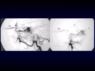
3D CE MP-RAGE • Flow gap seen in the left transverse sinus on 2D-TOF whereas CE MP-RAGE clearly demonstrates intravascular enhancement of a hypoplastic vessel • Patent venous sinus is confirmed on venous phase DSA 6. Liang et al.
3D CE MP-RAGE • MP-RAGE is also useful for direct visualization of thrombus in conjuction with other sequences • Apparent flow gap in 2D-TOF image is indeterminate, but clearly demonstrates thrombosis on MP-RAGE 6. Liang et al.
Summary • DVST is a rare entity, but diagnostically important as early treatment is related to eventual outcomes • TOF and PC are non-contrast, flow related techniques with acquisition parameters and artifact profiles that are important to keep in mind during interpretation • 3D CE MP-RAGE is not flow dependent, and is potentially superior to TOF and PC, but requires gadolinium contrast
References • Leach, James L., et al. "Imaging of Cerebral Venous Thrombosis: Current Techniques, Spectrum of Findings, and Diagnostic Pitfalls 1." Radiographics 26.suppl_1 (2006): S19-S41. • Ayanzen, R. H., et al. "Cerebral MR venography: normal anatomy and potential diagnostic pitfalls." American Journal of Neuroradiology 21.1 (2000): 74-78. • Rollins, Nancy, et al. "Cerebral MR Venography in Children: Comparison of 2D Time-of-Flight and Gadolinium-enhanced 3D Gradient-Echo Techniques 1."Radiology 235.3 (2005): 1011-1017. • Lotz, Joachim, et al. "Cardiovascular Flow Measurement with Phase-Contrast MR Imaging: Basic Facts and Implementation 1." Radiographics 22.3 (2002): 651-671. • Fera, Francesco, et al. "Comparison of different MR venography techniques for detecting transverse sinus stenosis in idiopathic intracranial hypertension."Journal of neurology 252.9 (2005): 1021-1025. • Liang, Luxia, et al. "Evaluation of the intracranial dural sinuses with a 3D contrast-enhanced MP-RAGE sequence: prospective comparison with 2D-TOF MR venography and digital subtraction angiography." American journal of neuroradiology 22.3 (2001): 481-492. • Leach, James L., et al. "Imaging of Cerebral Venous Thrombosis: Current Techniques, Spectrum of Findings, and Diagnostic Pitfalls 1." Radiographics26.suppl_1 (2006): S19-S41.