Download
1 / 23
230 likes | 290 Views
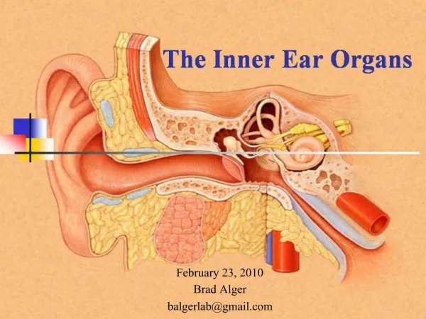
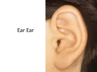
INNER EAR. General features. Ear has……………parts……….? Three parts External ear : collects sound waves Middle ear : transmits sound waves Internal ear : concerned with equilibration and hearing. Conduction of sound……………. Sound waves. Cochlear nerve.

E N D
General features Ear has……………parts……….? Three parts • External ear : collects sound waves • Middle ear : transmits sound waves • Internal ear : concerned with equilibration and hearing
Conduction of sound…………… Sound waves Cochlear nerve Air-conduction of sound
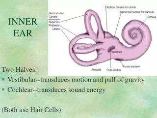
Internal ear General features • Lies within the petrous portion of temporal bone • Key contents of internal ear • Bony labyrinth contains perilymph • Membranous labyrinth is filled with endolymph and contains the sensory organs
Bony labyrinth • Cochlea • Vestibule • Bony semicircular canals
Cochlea • It resembles a snail’s shell • Consists of • Modiolus is the axial bony stem around which the cochlear canal spirals. It is the elongated cone. At base, it is perforated by the fibers of the cochlear nerve. • Cochlear spiral canal is spirally arranged around the modiolus. The basal turn bulges into the medial wall of middle ear cavity as promontory. Modiolus Cochlear spiral canal
Osseous spiral lamina • Osseous spiral lamina it is the spiral ridge of bone which projects from the modiolus into the cochlear canal. • The free edge splits into upper & lower lips seperated by C-shaped sulcus called spiralis internus Vestibular membrane & Basilar membrane • From the upper lip, the vestibular membrane extends to the outer wall o the cochlear canal • From the lower lip, the basilar membrane extends to the outer wall of the cochlear canal
Scala media (cochlear duct) • It is triangular thus enclosed by the vestibular & basal membranes and outer wall of the cochlear canal. Scala vestibuli & Scala tympani • The spiral lamina divides into scala vestibuli above & scala tympani below. • Both communicates with each other at the apex of the cochlea by small opening called helicotrema. scala vestibuli scalar media scala tympani
At the basal turn of cochlea……. The scala vestibuli communicates with anterior wall of vestibule. The scala tympani presents 2 features - fenestra cochleae (round window) :- it is closed by secondary tympanic membrane - beginning of the aqueduct of cochlea :- it is the narrow tubular canal through which perilymph within the cochlea communicates with the CSF of subarachnoid space.
Vestibule • Organ of balance • Hollow bony space present b/w cochleaanteriorly & semicircular canals posteriorly. It has
Ellipticalrecess Sphericalcalrecess Cochlear recess
Spiral recess & vestibular recess is seperated by the vestibular crest which splits inferiorly to enclose the cochlear recess.
Bony semicircular canals (anterior, posterior, and lateral) • Semicircular duct is present in each canal. • Canal at right angles to each other. • Dilated part of the canal is called as Ampulla.
Membranous labyrinth • Cochlear duct • Utricle • Saccule • Ampulla • Semicircular ducts
Cochlear duct Contains spinal organ (of Corti), the sound receptors lies on basilar membrane of cochlear duct
Utricle and saccule It detects the position of the head with respect to the gravity • Contain receptors - macula utricli and macula sacculi, which respond to linear acceleration and deceleration, static of gravity.
Semicircular ducts………… Evaluates the movement of the head & has special role in co-ordination of eye movements with movement of head. • Each duct has a membranous ampullae. • Containing crista ampullaris, receptors of balance that respond to rotational acceleration in three different planes
Labyrinthitis • Tinnitus • Meniere’s syndrome • Motion sickness • Nystagmus • Sensorineural deafness • Acoustic neuroma