Download
1 / 68
680 likes | 686 Views
Chapter 12: Physiology of the Muscular System. Anatomy & Physiology. Introduction. Muscular system is responsible for moving the framework of the body In addition to movement, muscle tissue performs various other functions. 2. General Functions.

E N D
Chapter 12: Physiology of the Muscular System Anatomy & Physiology
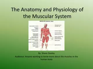
Introduction • Muscular system is responsible for moving the framework of the body • In addition to movement, muscle tissue performs various other functions 2
General Functions • Movement of the body as a whole or movement of its parts • Heat production • Posture 3
Function of Skeletal Muscle Tissue • Characteristics of skeletal muscle cells • Excitability (irritability)—ability to be stimulated • Contractility—ability to contract, or shorten, and produce body movement • Extensibility—ability to extend, or stretch, thereby allowing muscles to return to their resting length 4
Function of Skeletal Muscle Tissue • Overview of the muscle cell (Figures 12-1 and 12-2) • Muscle cells are called fibers because of their threadlike shape • Sarcolemma—plasma membrane of muscle fibers • Sarcoplasmic reticulum (SR) • T tubules—network of tubules and sacs found within muscle fibers • Membrane of the SR continually pumps calcium ions from the sarcoplasm and stores the ions within its sacs for later release (Figure 12-3) 5
Function of Skeletal Muscle Tissue • Overview of the muscle cell (cont) • Muscle fibers contain many mitochondria and several nuclei • Myofibrils—numerous fine fibers packed close together in sarcoplasm • Sarcomere • Segment of myofibril between two successive Z disks • Each myofibril consists of many sarcomeres • Contractile unit of muscle fibers 9
Function of Skeletal Muscle Tissue • Overview of the muscle cell (cont) • T tubules • Transverse tubules extend across the sarcoplasm at right angles to the long axis of the muscle fiber • Formed by inward extensions of the sarcolemma • Membrane has ion pumps that continually transport Ca++ ions inward from the sarcoplasm • Allow electrical impulses traveling along the sarcolemma to move deeper into the cell 10
Function of Skeletal Muscle Tissue • Overview of the muscle cell (cont) • Striated muscle (Figure 12-4) • Dark stripes called A bands; light H band runs across the midsection of each dark A band • Light stripes called I bands; dark Z disk extends across the center of each light I band • Triad • Triplet of tubules; a T tubule sandwiched between two sacs of sarcoplasmic reticulum • Allows an electrical impulse traveling along a T tubule to stimulate the membranes of adjacent sacs of the sarcoplasmic reticulum 11
Function of Skeletal Muscle Tissue • Myofilaments (Figures 12-5 and 12-6) • Each myofibril contains thousands of thick and thin myofilaments • Four different kinds of protein molecules make up myofilaments • Myosin • Makes up almost all the thick filament • Myosin “heads” are chemically attracted to actin molecules • Myosin “heads” are known as cross bridges when attached to actin 13
Function of Skeletal Muscle Tissue • Four different kinds of protein molecules make up myofilaments (cont) • Actin—globular protein that forms two fibrous strands twisted around each other to form the bulk of the thin filament • Tropomyosin—protein that blocks the active sites on actin molecules • Troponin—protein that holds tropomyosin molecules in place 16
Function of Skeletal Muscle Tissue • Mechanism of contraction • Excitation and contraction (Figures 12-7 through 12-13; Table 12-1) • A skeletal muscle fiber remains at rest until stimulated by a motor neuron • Neuromuscular junction—motor neurons connect to the sarcolemma at the motor endplate (Figure 12-7) • Neuromuscular junction is a synapse where neurotransmitter molecules transmit signals 17
Function of Skeletal Muscle Tissue • Excitation and contraction (cont) • Acetylcholine—the neurotransmitter released into the synaptic cleft that diffuses across the gap, stimulates the receptors, and initiates an impulse in the sarcolemma • Nerve impulse travels over the sarcolemma and inward along the T tubules, which triggers the release of calcium ions • Calcium binds to troponin, which causes tropomyosin to shift and expose active sites on actin 25
Function of Skeletal Muscle Tissue • Excitation and contraction (cont) • Sliding filament model (Figures 12-12 and 12-13) • When active sites on actin are exposed, myosin heads bind to them • Myosin heads bend and pull the thin filaments past them • Each head releases, binds to the next active site, and pulls again • The entire myofibril shortens 26
Function of Skeletal Muscle Tissue • Mechanism of contraction (cont) • Relaxation • Immediately after the Ca++ ions are released, the sarcoplasmic reticulum begins actively pumping them back into the sacs (Figure 12-3) • Ca++ ions are removed from the troponin molecules, thereby shutting down the contraction 27
Function of Skeletal Muscle Tissue • Mechanism of contraction (cont) • Energy sources for muscle contraction (Figure 12-14) • Hydrolysis of ATP yields the energy required for muscular contraction • ATP binds to the myosin head and then transfers its energy to the myosin head to perform the work of pulling the thin filament during contraction • Muscle fibers continually resynthesize ATP from the breakdown of creatine phosphate (CP) 28
Function of Skeletal Muscle Tissue • Energy sources for muscle contraction (cont) • Catabolic pathways • Aerobic pathway • Occurs when adequate O2 is available from blood (Figure 12-15) • Slower than anaerobic pathway, thus supplies energy for the long term rather than the short term 30
Function of Skeletal Muscle Tissue • Energy sources for muscle contraction (cont) • Catabolic pathways (cont) • Anaerobic pathway (Figure 12-16) • Very rapid, providing energy during first minutes of maximal exercise (Figure 12-16) • May occur when low levels of O2 are available • Results in the formation of lactic acid, which requires oxygen to convert back to glucose, the producing of an “oxygen debt” or excess postexercise oxygen consumption (EPOC) 32
Function of Skeletal Muscle Tissue • Energy sources for muscle contraction (cont) • Skeletal muscle contraction produces waste heat that can be used to help maintain the set point body temperature (Figure 12-17) 34
Function of Skeletal Muscle Organs • Muscles are composed of bundles of muscle fibers held together by fibrous connective tissue • Motor unit (Figure 12-18) • Motor unit—motor neuron plus the muscle fibers to which it attaches • Some motor units consist of only a few muscle fibers, whereas others consist of numerous fibers • Generally, the smaller the number of fibers in a motor unit, the more precise are the available movements; the larger the number of fibers in a motor unit, the more powerful the contraction available 36
Function of Skeletal Muscle Organs • Myography—method of graphing the changing tension of a muscle as it contracts (Figure 12-19) • Twitch contraction (Figure 12-20) • A quick jerk of a muscle that is produced as a result of a single, brief threshold stimulus (generally occurs only in experimental situations) 38
Function of Skeletal Muscle Organs • Twitch contraction (cont) • The twitch contraction has three phases • Latent phase—nerve impulse travels to the sarcoplasmic reticulum to trigger release of Ca++ • Contraction phase—Ca++ binds to troponin and sliding of filaments occurs • Relaxation phase—sliding of filaments ceases 41
Function of Skeletal Muscle Organs • Treppe—the staircase phenomenon (Figure 12-21, B) • Gradual, steplike increase in the strength of contraction that is seen in a series of twitch contractions that occur 1 second apart • Eventually, the muscle responds with less forceful contractions, and the relaxation phase becomes shorter • If the relaxation phase disappears completely, a contracture occurs 42
Function of Skeletal Muscle Organs • Tetanus—smooth, sustained contractions • Multiple wave summation—multiple twitch waves are added together to sustain muscle tension for a longer time • Incomplete tetanus—very short periods of relaxation occur between peaks of tension (Figure 12-21, C) 44
Function of Skeletal Muscle Organs • Tetanus—smooth, sustained contractions (cont) • Complete tetanus—the stimulation is such that twitch waves fuse into a single, sustained peak (Figure 12-21, D) • The availability of calcium determines whether a muscle will contract; if the calcium is continuously available, then contraction will be sustained (Figure 12-22) 45
Function of Skeletal Muscle Organs • Muscle tone • Tonic contraction—continual, partial contraction of a muscle • At any one time, a small number of muscle fibers within a muscle contract and produce a tightness or muscle tone • Muscles with less tone than normal are flaccid • Muscles with more tone than normal are spastic • Muscle tone is maintained by negative feedback mechanisms 47
Graded Strength Principle • Graded strength principle—skeletal muscles contract with varying degrees of strength at different times • Factors that contribute to the phenomenon of graded strength (Figure 12-26) • Metabolic condition of individual fibers • Number of muscle fibers contracting simultaneously; the greater the number of fibers contracting, the stronger the contraction • Number of motor units recruited • Intensity and frequency of stimulation (Figure 12-23) 48