Download
1 / 38
540 likes | 1.57k Views
IVIG Intravenous Immunoglobulin. Gabriela M a Claudia Tiago. IVIG ou IVIg Intravenous Immunoglobulin. IVIG is a blood product administered intravenously It contains the pooled Ig G extracted from the plasma of over one thousand blood donors

E N D
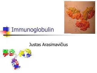
IVIG Intravenous Immunoglobulin Gabriela Ma Claudia Tiago
IVIG ou IVIg Intravenous Immunoglobulin • IVIG is a blood product administered intravenously • It contains the pooled IgG extracted from the plasma of over one thousand blood donors • Immunoglobulin products from human plasma were first used in 1952 to treat immune deficiency. It was initially shown to be effective in ITP in 1981. • Treatment for: Immune deficiencies (plasma protein replacement therapy) • Autoimmune Diseases (anti-inflammatory at a high dose ≈1-2 g/Kg) Acute Infections • IVIG cost is climbing and well over $50/g. ($8,000 for a 80 kg person at 2g/kg) • IVIG's effects last between 2 weeks and 3 months • primary immune dysfunction: 100 to 400 mg/kg of body weight every 3 to 4 weeks. • autoimmune diseases: 2 grams per kilogram of body weight for three to six months over a five day course once a month. Then maintenance therapy of 100 to 400 mg/kg of body weight every 3 to 4 weeks follows.
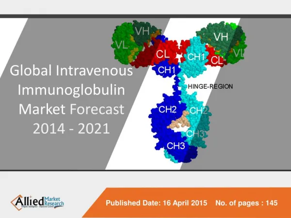
Rational basis • IVIG: pool of IgG • Immunoglobulins: effector molecules of immune defense • Igs properties: immune complex Diversity Specificity Variable domains Cellular effector pathways Constant domain Fc Fc
IVIG mechanism of action OK • Neutralization ??? Target: harmful antibodies • Idiotypic Network Theory A e B = IDIOTYPE A e C = ISOTYPE A C B Vaz & Pordeus. Visita à imunologia. Arq. Bras. Cardiol. vol.85 no.5 São Paulo Nov. 2005
Fc mechanism of IVIG action • Antibody Feedback: Cross-linkingbetween BCR andFcIIR B cell blocked
IVIG mechanism of action The precise mechanism by which IVIG suppresses harmful inflammation has not been definitively established BUT…. is believed to involve the Fc receptor… How? Which one(s)? FcαR FcαRI Fcα/μR FcεR FcεRI FcεRII FcγRI FcγRIIIA FcγR FcγRIIA FcγRIIB1 FcγRIIIB FcγRIIB2 FcRn
IVIG mechanism action • Fc Receptors (FcyR) • a protein found on the surface of NK cells, macrophages, neutrophils, mast cells and others • FcγRs are the most important Fc receptors for inducing phagocytosis of opsonized microbes
Glycoforms of IgG (Asn297) Carbohydrate whit terminal sugar residues such as galactose, sialic acid, N-acetylglucosamine, and fucose • more than 30 different antibody glycovariants have been detected in human serum, with about 25%–30% of them in the IgGglycoform. • Thus, these variants, multiplied by the four different IgG subclasses, result in more than 120 different glycoproteins in the IVIG preparation that could contain the active anti-inflammatory component
IVIG has anti-inflammatory effect at a high dose ≈1-2 g/Kg • 120 different glycoproteins in the IVIG preparation • terminal sugar residues of sialic acid confers anti-inflammatory property • 1-3% of IgGs in IVIG have sFc (sialylation) • recombinant sFc: enhanced 35 fold of action in vivo Carbohydrate Carbohydrate-Binding Proteins C-Type Lectins Siglecs Galectins CD1
DC-SIGN is a C-type lectin receptor • binds to mannose type carbohydrates. • Phagocytosis • Cell rolling interactions (ICAM) and activation of CD4+ T cells • Binds sFc anti-inflammatory responses • Population of regulatory macrophage • Splenic Marginal Zone
Maria Claudia Tiago
Objective: • To define the mechanism by which the 2,6-sialylated Fc mediates an anti- inflammatory response • To identify the properties of the regulatory macrophage population • To identify the receptor required for initiating this pathway in response to 2,6-sialylatedFc.
Results Are the splenic marginal zone macrophage necessary for IVIG-mediated immune suppression? 1 hourafter Clinical score analysis Arthritis inducing sera (K/BxN) IVIG Defined defects in specific immune cell populations Specificmacrophagepopulations in the splenic marginal zone might be required for the anti-inflammatory effect of the 2,6 sialylatedFcfound in IVIG
Results Which receptor expressed in macrophages is required for IVIG protection? 1 hourafter 1 hourafter Arthritis inducing sera (K/BxN) IVIG Blocking antibodies Interacting with glycopeptides: Scavenger receptor (MARCO) – bacterias Sialoadhesin receptor (CD169) – sialic acid C-type lectin receptor (SIGN-R1) – polysaccharide dextran TKO-SIGNR1 -antibody that results in the transient down-regulation of SIGN-R1 expression
Results Which receptor expressed in macrophages is required for IVIG protection? IVIG (2,6 Fc) 1 hourafter Clinical score analysis Arthritis inducing sera (K/BxN) C57BL/6 and SIGN-R1-/- Ankle bones The c-type lectin, SIGN-R1, is required for IVIG protection
Results Did SIGN-R1 able to bind to the 2,6-sialyted Fc? Pulsed with flourochrome-labeded 2,6-Fcs (red) Transfected macrophage (RAW-SIGN-R1) SIGN-R1 binds2,6-sialylatedFc.
Results Did SIGN-R1 able to bind to the 2,6-sialyted Fc and asialylated Fcs? The amount of bound Fcs were determined C57BL/6 mice Lack all IgG Fc receptors SIGN-R1-/- Resident peritoneal m were harvested Pulsed with 2,6-Fcs or asialylated Fcs The 2,6-sialylation of the IgGFc converts the molecule to a species that acquires the ability to engage a mSIGN-R1 and mediate an antiinflammatoryresponse.
Results Human DC-SIGN expressed on dendritic cells Yellow – Identical amino acids Green – Similar amino acids CRD- carbohydrate recognition domains
Results Did DC-SIGN able to bind to the 2,6-sialyted Fc? CHO cells expressing SIGN-R1, hDC-SIGN or hFcRIIb Pulsed with 2,6-Fcs Mannan = ligand for DC-SIGN Fibrinogen = similar to Fc linked glycan Human DC-SIGN, binds 2,6-sialylatedFc
Results 2,6-sialylation Fc 1 hourafter 1 hourafter Arthritis inducing sera (K/BxN) C57Bl/6 SIGN-R1-/- FcγRIIb-/- IVIG FcR binding mSIGN-R1, hDC-SIGN binding antiinflammatory response FcRIIb
Results Did FcRIIb involve in the mechanism by which the 2,6-Fc mediates an anti-inflammatory response? K/BxN The absence of FcRIIb in the recipient prevented the protection afforded by these splenocytes
Objective: To study hDC-SIGN in the context of IVIG anti-inflammatory activity in expressing-hDC-SIGN mice.
Could hDC-SIGN mediate anti-inflammatory protection by IVIG? WT SIGN-R1-/- hDC-SIGN+/SIGN-R1-/- Treated with sFc Challenged with arthritogenic K/BxN serum Clinical score assessement hDC-SIGN substitutes for SIGN-R1 in mediating IVIG anti-inflammatory protection
Were hDC-SIGN+ macrophages sufficient to induce an anti-inflammatory response? WT hDC-SIGN+ BMMФ + 30min sFc or asyaloFc Transfered to WT mice WT Challenged with K/BxN Clinical score assessement hDC-SIGN+ Macrophages treated with sFC showed reduced joint inflammation
Is FcγRIIB required to the anti-inflammatory property induced hDC-SIGN+ macrophages? hDC-SIGN+-BMMФ + 30min hDC-SIGN+ sFc or PBS Transfered to FcγRIIB-/- SIGN-R1-/- Challenged with K/BxN Clinical score assessement The anti-inflammatory property induced by hDC-SIGN+ macrophages depends on FcγRIIB
Was IL-4 responsable for mediating IVIG anti-inflammatory activity? hDC-SIGN+ BMMФ + 30min sFc or PBS Transfered to IL-4-/- WT Challenged with K/BxN Clinical score assessement IL-4 is crucial for mediating IVIG anti-inflammatory activity
Could Th2 cytokines supress K/BxN-induced inflammation? Treated with IL-4, IL-13 or IL-3 FcγRIIB-/- WT K/BxN Clinical score evaluation Inflammation was attenuated after Th2 cytokines administration
Did sFc administration increase Th2 cytokines production? WT SIGN-R1-/- Treated with sFc (1h) Splenic cells were removed Quantification of IL-4, IL-33 and IL-25 mRNA expression (qPCR) IL-33 mRNA was upregulated in WT mice after sFc administration
Can IL-33 induce IL-4 production? Treated with PBS, IL-33, IL-25 or TSLP WT K/BxN Clinical score evaluation and analyses of IL-4 levels IL-33 reverts K/BxN-induced inflammation by increasing IL-4 levels
Does Anti-IL-33Rα ablate the sFc protection? Treated with sFc or sFc+anti-IL-33Rα hDC-SIGN+/SIGN-R1-/- K/BxN Clinical score evaluation The IL-33Rα blocking increases joint inflammation
Did IL-33 and IL-4 increase FcγRIIB expression on monocytes? hDC-SIGN+-Monocytes (CD11b+Ly6G+) + PBS or IL-4 or IL-33 or IL-25 24h FcγRIIB expression by FACS FcγRIIB expression on monocytes was increased after IL-33 and IL-4 treatment
Are basophils involved with reduced joint inflammation? Treated with sFc or sFc+anti-FcεRI hDC-SIGN+/SIGN-R1-/- K/BxN Clinical score evaluation Basophils contribute for IVIG anti-inflammatory activity
Are basophils the main source of IL-4 production during sFc treatment? Treated with PBS or sFc IL-4-GFP mice K/BxN Clinical score evaluation and quantification of IL-4-producing basophils Increased IL-4-producing basophils were induced during sFC treatment
Were basophils associated with anti-inflammatory activity induced by sFc? WT or FcγRIIB-/-Basophils (DX5+FcεRI+c-Kit-) + PBS, IVIG or IL-33 Transfered to WT Challenged with K/BxN IL-33-treated basophils also increased anti-inflammatory activity in a FcγRIIB-dependent manner


![IS THERE STILL ANY ROLE FOR THE USE OF INTRAVENOUS IMMUNOGLOBULIN [IVIG] IN RECURRENT SPONTANEOUS MISCARRIAGE [RSM]?](https://cdn0.slideserve.com/1456233/slide1-dt.jpg)