Download
1 / 30
300 likes | 523 Views
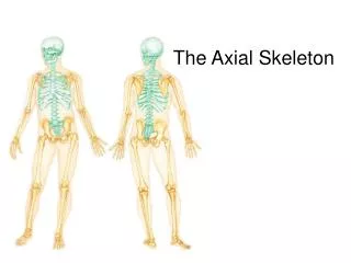
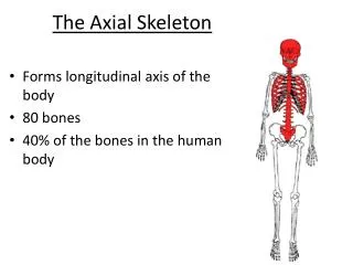
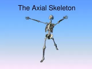
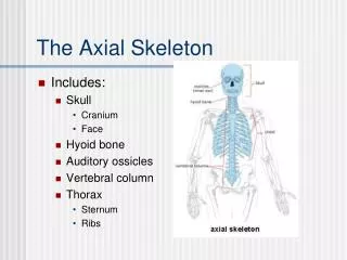
The Axial Skeleton. Forms the longitudinal axis of the body Divided into three parts Skull Vertebral column Bony thorax. The Axial Skeleton. Figure 5.6a. The Axial Skeleton. Figure 5.6b. The Skull. Two sets of bones Cranium Facial bones Bones are joined by sutures

E N D
The Axial Skeleton • Forms the longitudinal axis of the body • Divided into three parts • Skull • Vertebral column • Bony thorax
The Axial Skeleton Figure 5.6a
The Axial Skeleton Figure 5.6b
The Skull • Two sets of bones • Cranium • Facial bones • Bones are joined by sutures • Only the mandible is attached by a freely movable joint
Paranasal Sinuses • Hollow portions of bones surrounding the nasal cavity • Functions of paranasal sinuses • Lighten the skull • Give resonance and amplification to voice
Paranasal Sinuses Figure 5.10a
Paranasal Sinuses Figure 5.10b
The Hyoid Bone • The only bone that does not articulate with another bone • Serves as a moveable base for the tongue • Aids in swallowing and speech
The Hyoid Bone Figure 5.12
The Fetal Skull • The fetal skull is large compared to the infant’s total body length • Fontanels—fibrous membranes connecting the cranial bones • Allow the brain to grow • Convert to bone within 24 months after birth
The Fetal Skull Figure 5.13a
The Fetal Skull Figure 5.13b
The Vertebral Column • Each vertebrae is given a name according to its location • There are 24 single vertebral bones separated by intervertebral discs • Seven cervical vertebrae are in the neck • Twelve thoracic vertebrae are in the chest region • Five lumbar vertebrae are associated with the lower back
The Vertebral Column • Nine vertebrae fuse to form two composite bones • Sacrum • Coccyx
The Vertebral Column Figure 5.14
The Vertebral Column • The spine has a normal curvature • Primary curvatures are the spinal curvatures of the thoracic and sacral regions • Present from birth • Secondary curvatures are the spinal curvatures of the cervical and lumbar regions • Develop after birth
The Vertebral Column Figure 5.15
The Vertebral Column Figure 5.16
Sacrum and Coccyx • Sacrum • Formed by the fusion of five vertebrae • Coccyx • Formed from the fusion of three to five vertebrae • “Tailbone,” or remnant of a tail that other vertebrates have
Sacrum and Coccyx Figure 5.19
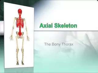
The Bony Thorax • Forms a cage to protect major organs • Consists of three parts • Sternum • Ribs • True ribs (pairs 1–7) • False ribs (pairs 8–12) • Floating ribs (pairs 11–12) • Thoracic vertebrae
The Appendicular Skeleton • Composed of 126 bones • Limbs (appendages) • Pectoral girdle • Pelvic girdle
The Appendicular Skeleton Figure 5.6a
The Appendicular Skeleton Figure 5.6b
The Pectoral (Shoulder) Girdle • Composed of two bones • Clavicle—collarbone • Scapula—shoulder blade • These bones allow the upper limb to have exceptionally free movement
Bones of the Shoulder Girdle Figure 5.21a
Gender Differences of the Pelvis • The female inlet is larger and more circular • The female pelvis as a whole is shallower, and the bones are lighter and thinner • The female ilia flare more laterally • The female sacrum is shorter and less curved • The female ischial spines are shorter and farther apart; thus the outlet is larger • The female pubic arch is more rounded because the angle of the pubic arch is greater
Gender Differences of the Pelvis Figure 5.24c
Arches of the Foot • Bones of the foot are arranged to form three strong arches • Two longitudinal • One transverse
Arches of the Foot Figure 5.27