Download
1 / 57
580 likes | 856 Views
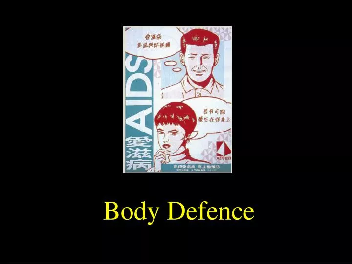
Body Defence. BODY DEFENCE MECHANISMS The body defends itself against physical injuries and invasion by harmful materials and organisms in various ways. These ways can be divided into PREVENTION and CURE . 1. PREVENTION. secretions. mechanical barriers. 1. PREVENTION

E N D
BODY DEFENCE MECHANISMS • The body defends itself against physical injuries and invasion by harmful materials and organisms in various ways. • These ways can be divided into PREVENTION and • CURE.
1. PREVENTION secretions mechanical barriers
1. PREVENTION • (a) Against physical injuries • (i)Tough outer coating • - keratinised compound squamous epithelium of skin. • The skin is thickest in areas where physical injury is most common • e.g. palms of hands, soles of feet. • (ii) Adipose tissue • - forms a cushion between skin and underlying organs. • Some delicate organs, e.g. kidneys, may be further protected by a coat of fat. • (iii) Bones • - the delicate haemopoietic (blood forming) tissues are encased in the shafts of long bones of the limbs and the sternum (red bone marrow). • The brain, spinal cord, heart and lungs are also protected by bones.
(b) Against invasion by harmful materials and organisms • (i) Intact skin • - relatively few chemicals and organisms are able to penetrate intact skin. • (ii) Cilia • - beating of cilia on the outer surface of epithelial cells in the respiratory tract he1ps to prevent harmful dust/bacteria etc. reaching the lungs. • (iii) Secretions • - many body secretions contain chemicals which are harmful to many pathogenic organisms. • e.g. gastric juice (pH 2) • tears (from lachrymal glands) • mucus - respiratory tract, vagina • sebaceous secretions • lysozyme in tissue fluid
2 CURE • There are two important mechanisms by which the body can remove harmful materials / organisms once they have entered the body. • These mechanisms are: • I. INFLAMMATORY RESPONSE • II. SPECIFIC IMMUNE RESPONSE
The inflammatory response is a local response to tissue damage and invasion by harmful materials and/or organisms. • The response is often described as being “non-specific” as the response is much the same regardless of the nature of the issue damage or foreign material/ organisms. • The strength of an inflammatory response does, however, vary according to the severity of an injury. • The inflammatory response can: • - remove dead and damaged body cells • - remove harmful materials and cells
Specific immune responsesdiffer from inflammatory responses in that an immune response is a ‘specific’ response to invasion by harmful materials and/or cells. • Thus a particular immune response will deal specifically with the material or organism which stimulated the response. • Immune responses cannot deal with dead or damaged body cells, however certain cells of the immune system can ‘remember’ a particular harmful material or organism and can react very quickly to a second or subsequent invasion by that substance.
I. INFLAMMATORY RESPONSE Events during localized infection (a non-specific mechanism): permeability, blood flow, vasodilation Histamine is released from mast cells WBCs emerge from blood vessel
I. INFLAMMATORY RESPONSE • When connective tissues and blood vessel walls are damaged by physical injury or the presence of harmful materials/cells, certain cells respond by liberating a variety of chemicals. • These chemicals have two major functions: • (a) Capillary dilation, • resulting in increased blood flow in the damage area. • (b) Increase in capillary permeability, allowing blood plasma and neutrophil phagocytes to pass into the surrounding fluid. • (a) and (b) cause the inflamed area to become: • (i) red • (ii) swollen • (iii) warmer than surrounding tissues • (iv) painful (due to pressure of increased fluid on local endings)
The phagocytes which have migrated from blood vessels engulf dead and damaged cells, thus cleaning the wound, and also phagocytose harmful materials and organisms. • Once cell debris and ‘foreign’ materials are removed tissue repair can take place. • The inflammatory response is not always sufficient to destroy and remove harmful materials and organisms, and these may migrate from the site of injury to other parts of the body via the blood and lymph. • It is in such situations that a specific immune response occurs.
To lymph node To spleen
II SPECIFIC IMMUNE RESPONSE • Lymphocytes are the most important cells in immune responses, although other cells are also involved, such as macrophages. • Lymphocytes are found in the blood (20-25% of white blood cells) and in lymphoid organs such as the bone marrow, thymus, lymph nodes (often called glands) and spleen. • Any substance which is recognized as ‘foreign’ by the body and which can stimulate an immune response is called an ANTIGEN (Ag). • Antigens may be soluble macromolecules, cell surface components or chemicals synthesized by foreign cells.
Commonly encountered antigens include: • Bacteria ( components of cell walls and flagellae, toxins) • Viruses ( protein ‘coat’ subunits) • Fungi and protozoa ( cell surface components) • Macromolecules ( especially proteins) • Less frequently encountered antigens include: • RBC’s (‘blood group substances’ on membranes) • grafted cells (cell membrane proteins or glyco-proteins)
There are two kinds of immune responses: • - ANTIBODY response (or humoral immune response) • - CELLULAR response (or cell-mediated immune response) • In general, ANTIBODY responses deal with bacterial and RBC antigens and possibly fungal and protozoal antigens, • whilst the CELLULAR response deals with viral antigens, grafted cells and possibly fungal and protozoal antigens.
(a) Development of immune responsiveness • (i) Lymphocytes develop in the bone marrow from haemopoietic precursor cells. • (ii) Some lymphocytes nature fully in the bone marrow to become B lymphocytes (B cells). • (iii)Other, immature, lymphocytes pass from the bone marrow to the thymus where they mature into T lymphocytes (T cells). • (iv)Both B and T cells pass to the lymph nodes and spleen via the blood stream. • (N.B. Lymph nodes and spleen can be regarded as a complex organization of three types of cell involved in the initiation of the immune reaction - lymphocytes, plasma cells and phagocytic cells of the mono-nuclear phagocyte system.
Stem cell leucocytes • Different types of white blood cells Granulocytes (polymorphonuclear Leucocytes-PMN) agranulocytes lymphocytes T cell B cell Basophil 0-1% Neutrophile 50-70% phagocytic RBS Eosinophil 1-4% Allergic? Monocyte 2-8% platelets 33% erythrocytes Macrophage phagocytic Mast cell Releases histamine
Different types of white blood cells • 1) Granulocytes (polymorphonuclear leucocytes or cells) • - White blood cells that possess GRANULES in their cytoplasm, with nuclei & a few days of life span, • - a) Neutrophils: • - the most abundant PMN, an important PHAGOCYTIC cell for NON-SPECIFIC body defence; • - amoeboid, can leave blood vessels & enter into tissue. • b) Eosinophils: • quite rare, functions uncertain but probably phagocytic & associated with hypersensitivity & allergic reactions. • c) Basophils: • - smallest number, NON-PHAGOCYTIC but becomes MAST CELLS when entered tissues; • - contains HISTAMINES which when released, will cause vaso-dilation, increased blood flow, increased permeability of blood vessels & outflow of cells.
2) Agranulocytes (no granules in the cytoplasm) • a) lymphocytes (33%): • B CELLS & T CELLS which are small cells with large nucleus; • b) monocytes (2-8%): • Phagocytic, becomes macrophage in tissues & have kidney-shaped nucleus
**Development of B & T cells • Both B & T cells originate from a stem cell in haemopoietic tissues (yolk sac & liver in foetus; bone marrow in adults). • Some migrate via blood to the thymus & develop into T cells/lymphocytes. • These T cells then migrate to lymph nodes & spleens where most of them reside and be ready for specific immure responses. • Thymus, being an important organ for the differentiation of stem cells into specific lymphocytes, is called the PRIMARY LYMPHOID TISSUE while lymph nodes & spleen where the mature immunocompetent cells lie are called SECONDARY LYMPHOID TISSUES
In birds, some stem cells migrate from haemopoietic tissues to an organ called Bursa of Fabricius and develop into B cells/lymphocytes. • The B cells then migrate to the lymph nodes & spleen and reside there for specific immune responses. • The bursa equivalent for mammals is unknown. The bone marrow is thought to be a likely place. • When B & T cells arrive at the secondary lymphoid organs (lymph nodes & spleen), they settle in separate specific areas. • There are specific T & B areas in these organs.
Primary lymphoid tissues Secondary lymphoid tissues /spleen
The secondary lymphoid organs are the places where B & T cells accumulate. • It is also the place where pathogens in blood & lymph are caught. • It is in these places that pathogens stimulate the B & T cells and TURN ON specific immune responses. • Most specific responses take place at these sites. They are therefore the battle grounds for specific mechanisms!
**antigen processing ** T cell response or B cell response
STIMULATION OF SPECIFIC RESPONSES • When pathogens reach the lymph node or spleen, they may be first processed by the macrophages at these sites (antigen processing). • The processed antigens may then stimulate either the T or B cells (or both in some cases) and turn on the CMIR or HIR respectively. • Sometimes, pathogens need not go through “antigen processing” and can stimulate B and T cells directly.
(b) Reaction of lymphocytes to antigen • (i) In the lymph nodes or spleen the antigen stimulates B and / or T lymphocytes as follows: • (ii) ( I ) Humoral response:
antigen Memory cell Blast cell antibodies OR T-helper cell for T-dependent antigens Antibody forming cell Plasma cell
Primary immune response Secondary immune response antigen B cell Same antigen Blast cell Antibody forming cell Plasma cell which produce antibodies Differentiation of B cells
The characteristics of the humoral response is that B cells are involved & the process results in the production of ANTIBODIES specific for the antigen. • THE PRIMARY RESPONSE (elicited when an antigen entered into the body for the FIRST TIME): • When an antigen reaches the lymph node/spleen, it will stimulate the appropriate B cell there which is specific to it. • Some antigens cannot turn on the B cell directly, they need the presence of T cells & these antigens are called T-dependent antigens (in contrast to T-independent antigens).
The stimulated B cell will then differentiate & multiply into BLAST CELLS. • These in turn proliferate & differentiate into ANTI-BODY FORMING CELLS and then plasma cells which are very efficient in producing ANTI-BODIES which can then act on the antigen.
Receptor sites for antigens • The antibodies are Y-shaped structures which are also called IMMJJNOGLOBULINS as they are protein molecules. • The top ends of the “Y” are specific to the particular antigen & can therefore bind to it. High affinity for PMNs, macrophage
The antibodies can help to destroy antigens in three main ways: • 1) Bacterial lysis by antibody
bacterium antibodies Attachment of antibodies Complement attached to bacterium-antibody complex A hole is drilled by the complex, resulting in lysis Lysis and death
The antibodies can help to destroy antigens in three main ways: • 1) Bacterial lysis by antibody 2) Enhanced phagocytosis
Enhanced phagocytosis: coat Bacterium is “slippery” Phagocyte cannot grasp it bacterium Antibodies attached to coat of bacterium Since end of antibody has high affinity for phagocyte, phagocyte can now grasp bacterium through antibody
The antibodies can help to destroy antigens in three main ways: • 1) Bacterial lysis by antibody • 2) Enhanced phagocytosis 3) Certain antibodies can neutralize bacterial toxins by forming antigen-antibody complexes which are then phagocytosed. Eosinophils are especially active in phagocytosing antigen-antibody complexes.
SECONDARY RESPONSE: • Memory B lymphocytes are long-lived cells which remain dormant in lymphoid tissues for many months or years, until the antigen which stimulated their production is encountered again. • If and when this occurs the memory B Lymphocytes respond immediately to antigen by dividing and differentiating into more plasma cells and memory B lymphocytes.
Stronger and faster Latent period Latent period Re-injection 1st injection Secondary response Primary response
Compared with the original, or primary response toantigen the secondary immune response is: • - larger • - faster • - longer lasting, as once stimulated, the memory cells continue to divide for monthsor years.
(II)Cellular response • T lymphocytes • cell divisions and differentiation • cytotoxic T lymphocytes • and + ‘memory’ T lymphocytes • activated T lymphocytes
The cell-mediated immune response (CMIR) Memory cell Killer T cell (cytotoxic T cell) Kill antigen directly lymphokines antigen Activated T cell *No antibodies are involved Activated macrophage kills antigen monocyte macrophage
Cytotoxic T lymphocytes • these migrate (if necessary) to the site of the antigen, which is normally a cell with antigen on its surface. • The cytotoxic T cells are capable of killing cells with antigen on their surfaces. • These ‘target’ cells then lyse and the fragments are phagocytosed. • As ‘in the antibody response, cytotoxic T cells have a particular specificity, only reacting with cells bearing the antigen which stimulated their production.
Activated T lymphocytes • - these liberate chemicals called lymphokines. • The lymphokines then activate the macrophages into activated macrophages which are highly efficient in eating and killing the antigens. • Memory T lymphocytes are very similar to memory B lymphocytes, except that subsequent stimulation by antigen results in rapid production of more cytotoxic T lymphocytes and memory T lymphocytes. • The same graph showing primary and secondary responses applies except that the y axis becomes ‘number of cytotoxic T lymphocytes’.
(I) IMMUNITY ANY IHM1UNITSATOIN • Immunity of the body refers to “all those physiologic mechanisms that endow the animal with the capacity to recognize materials as foreign to itself and to neutralize, eliminate, or metabolize them with or without injury to itsown tissues”. • Immunity to a disease may be acquired naturally or artificially.
Natural Immunity • 1. Natural passive immunity is important in young babies when antibodies in the mother’s blood diffuse across the placenta before birth. • Maternal antibodies are also present in breast milk. • As antibodies are catabolised in a few months the protective effect is short-lived. • 2. Natural active immunity results from infection witha pathogen, resulting in the individual producing his own antibodies etc. • If the disease has a long incubation period then both primary and secondary responses occur, resulting long-term immunity, e.g. mumps, chicken pox.
Artificial Immunity • 1. Artificial passive immunity involves an injection of ‘ready-made’ antibodies, usually obtained from animals immunised with the antigen. • The immunity is short-lived (3-6 months) as the antibodies are catabolised, however this is a good method of immunisation if instant immunity is required.
2. Artificial active immunity involves the introduction of killed or non-virulent strains of disease-causing organisms into the body. • Humoral and/or cellular responses then take place. • Generally at least two doses of antigen are given atsuitable intervals, so that the secondary immune response is stimulated.
There are three main types of bacterial and viral antigen preparations in current use: • (a) Toxoids • Soluble toxins of bacteria such as diphtheria and tetanus, modified and made less toxic by adding formalin or gentle-beating. • Treatment destroys the toxic parts of the antigen molecule, leaving the antigenic sites unchanged. • (b) Killed organisms • Cultured organisms killed by heat, UV light or chemicals, e.g.whooping cough (pertussis), poliomyelitis (Salk), cholera,typhoid.
(c) Live, “attenuated” organisms • Vaccines made from strains of organisms that have lost their virulence. • The emergence of an attenuated strain is a combination of science and luck • e.g. BCG - a virulent strain of Mycobacterium tuberculosis was grown in a medium containing bile salts which resulted in the production of an attenuated strain - Bacillus Calmette - Guerin (1908). • Other examples of vaccines prepared from attenuated organisms are poliomyelitis (Sabin), measles, rubella, yellow fever.