Download
1 / 47
620 likes | 1.83k Views
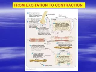
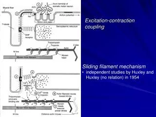
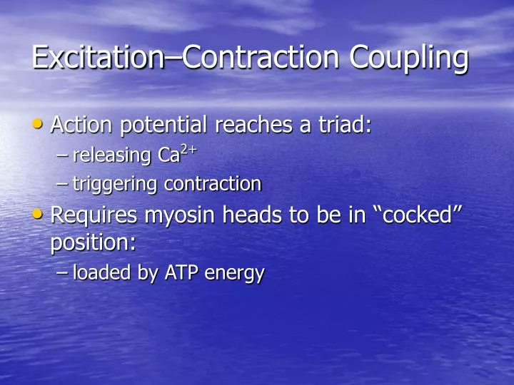
Excitation–Contraction Coupling. Action potential reaches a triad: releasing Ca 2+ triggering contraction Requires myosin heads to be in “cocked” position: loaded by ATP energy. Exposing the Active Site. Figure 10–11. The Contraction Cycle. Figure 10–12 (1 of 4). The Contraction Cycle.

E N D
Excitation–Contraction Coupling • Action potential reaches a triad: • releasing Ca2+ • triggering contraction • Requires myosin heads to be in “cocked” position: • loaded by ATP energy
Exposing the Active Site Figure 10–11
The Contraction Cycle Figure 10–12 (1 of 4)
The Contraction Cycle Figure 10–12 (2 of 4)
The Contraction Cycle Figure 10–12 (3 of 4)
The Contraction Cycle Figure 10–12 (Navigator) (4 of 4)
5 Steps of the Contraction Cycle • Exposure of active sites • Formation of cross-bridges • Pivoting of myosin heads • Detachment of cross-bridges • Reactivation of myosin
Fiber Shortening • As sarcomeres shorten, muscle pulls together, producing tension Figure 10–13
Contraction Duration • Depends on: • duration of neural stimulus • number of free calcium ions in sarcoplasm • availability of ATP
Relaxation • Ca2+ concentrations fall • Ca2+ detaches from troponin • Active sites are recovered by tropomyosin • Sarcomeres remain contracted
A Review of Muscle Contraction Table 10–1 (1 of 2)
KEY CONCEPT(Part 1) • Skeletal muscle fibers shorten as thin filaments slide between thick filaments • Free Ca2+ in the sarcoplasm triggers contraction • SR releases Ca2+ when a motor neuron stimulates the muscle fiber
KEY CONCEPT(Part 2) • Contraction is an active process • Relaxation and return to resting length is passive
Tension Production • The all–or–none principal: • as a whole, a muscle fiber is either contracted or relaxed
Tension of a Single Muscle Fiber • Depends on: • the number of pivoting cross-bridges • the fiber’s resting length at the time of stimulation • the frequency of stimulation
Frequency of Stimulation • A single neural stimulation produces: • a single contraction or twitch • which lasts about 7–100 msec • Sustained muscular contractions: • require many repeated stimuli
3 Phases of Twitch • Latent period before contraction: • the action potential moves through sarcolemma • causing Ca2+ release
3 Phases of Twitch • Contraction phase: • calcium ions bind • tension builds to peak
3 Phases of Twitch • Relaxation phase: • Ca2+ levels fall • active sites are covered • tension falls to resting levels
Treppe • Repeated stimulations immediately after relaxation phase: • stimulus frequency < 50/second • Causes a series of contractions with increasing tension
Wave Summation • Increasing tension or summation of twitches Figure 10–16b
Incomplete Tetanus • Twitches reach maximum tension Figure 10–16c
Complete Tetanus Figure 10–16d
Muscle Tone • The normal tension and firmness of a muscle at rest • Muscle units actively maintain body position, without motion • Increasing muscle tone increases metabolic energy used, even at rest
2 Types of Skeletal Muscle Tension • Isotonic contraction • Isometric contraction
Isotonic Contraction Figure 10–18a, b
Isometric Contraction Figure 10–18c, d
ATP and Muscle Contraction • Sustained muscle contraction uses a lot of ATP energy • Muscles store enough energy to start contraction • Muscle fibers must manufacture more ATP as needed
ATP and CP Reserves • Adenosine triphosphate(ATP): • the active energy molecule • Creatine phosphate(CP): • the storage molecule for excess ATP energy in resting muscle
ATP Generation • Cells produce ATP in 2 ways: • aerobic metabolism of fatty acids in the mitochondria • anaerobic glycolysis in the cytoplasm
Aerobic Metabolism • Is the primary energy source of resting muscles • Breaks down fatty acids • Produces 34 ATP molecules per glucose molecule
Anaerobic Glycolysis • Is the primary energy source for peak muscular activity • Produces 2 ATP molecules per molecule of glucose • Breaks down glucose from glycogen stored in skeletal muscles
Energy Use and Muscle Activity • At peak exertion: • muscles lack oxygen to support mitochondria • muscles rely on glycolysis for ATP • pyruvic acid builds up, is converted to lactic acid
Results of Muscle Fatigue • Depletion of metabolic reserves • Damage to sarcolemma and sarcoplasmic reticulum • Low pH (lactic acid) • Muscle exhaustion and pain
The Recovery Period • The time required after exertion for muscles to return to normal • Oxygen becomes available • Mitochondrial activity resumes
Oxygen Debt • After exercise: • the body needs more oxygen than usual to normalize metabolic activities • resulting in heavy breathing
Hormones and Muscle Metabolism • Growth hormone • Testosterone • Thyroid hormones • Epinephrine
Structure of Cardiac Tissue • Cardiac muscle is striated, found only in the heart Figure 10–22
Intercalated Discs • Are specialized contact points between cardiocytes • Join cell membranes of adjacent cardiocytes (gap junctions, desmosomes)
Functions of Intercalated Discs • Maintain structure • Enhance molecular and electrical connections • Conduct action potentials
Smooth Muscle in Body Systems (Part 1) • Forms around other tissues • In blood vessels: • regulates blood pressure and flow • In reproductive and glandular systems: • produces movements
Smooth Muscle in Body Systems (Part 2) • In digestive and urinary systems: • forms sphincters • produces contractions • In integumentary system: • arrector pili muscles cause goose bumps
Structure of Smooth Muscle • Nonstriated tissue Figure 10–23
Comparing Smooth and Striated Muscle • Different internal organization of actin and myosin • Different functional characteristics
8 Characteristics of Smooth Muscle Cells • Long, slender, and spindle shaped • Have a single, central nucleus • Have no T tubules, myofibrils, or sarcomeres • Have no tendons or aponeuroses
8 Characteristics of Smooth Muscle Cells • Have scattered myosin fibers • Myosin fibers have more heads per thick filament • Have thin filaments attached to dense bodies • Dense bodies transmit contractions from cell to cell
Functional Characteristics of Smooth Muscle • Excitation–contraction coupling • Length–tension relationships • Control of contractions • Smooth muscle tone