Download
1 / 30
300 likes | 307 Views
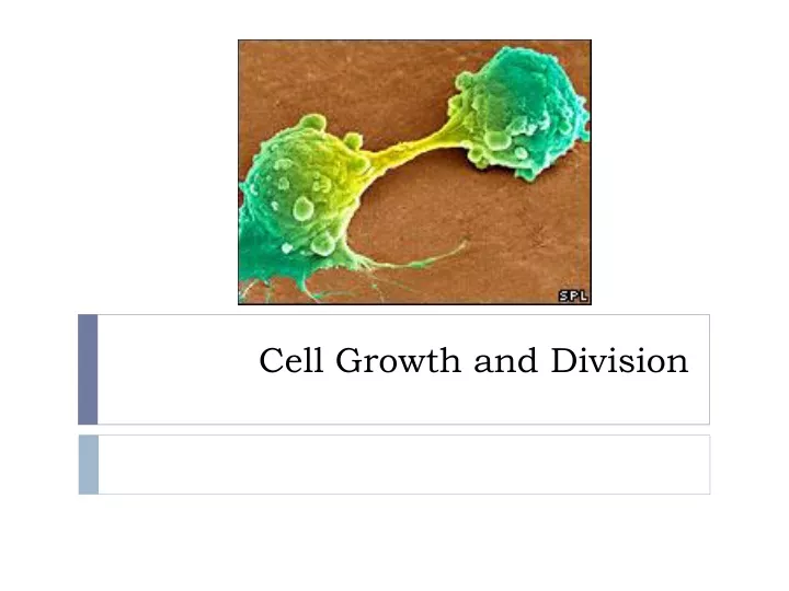
Cell Growth and Division. The Cell Cycle. The cycle of growth, DNA synthesis, and division is essential for an organism to grow and heal. If it goes out of control, abnormal cell growth may occur, resulting in cancer cells. Aveoli in the lung. Lung cancer cell. The Cell Cycle.

E N D
The Cell Cycle • The cycle of growth, DNA synthesis, and division is essential for an organism to grow and heal. • If it goes out of control, abnormal cell growth may occur, resulting in cancer cells. Aveoli in the lung Lung cancer cell
The Cell Cycle • The cell cycle is the regular pattern of growth, DNA duplication, and cell division that occurs in eukaryotic cells.
Cell Cycle: Four Main Stages • The four main stages of the cell cycle are: • Gap 1 • Synthesis • Gap 2 • Gap 1, Synthesis, Gap 2 = Interphase • Mitosis • Scientists were limited by the microscopes of the time and named the “gaps” because they weren’t able to see activity in the cell at these times.
Gap 1 (G1) • During G1, the cell carries out its normal functions. • The cell will increase in size, and its organelles will increase in number. • A cell spends most of it’s time in this stage, but the length of the stage depends on the cell type. • This is the stage where the cell determines if it will undergo apoptosis, or divide.
Synthesis (S) • The cell makes a copy of its nuclear DNA during this stage. • By the end of this stage, the cell contains two complete sets of DNA. DNA as seen from an SEM
Gap 2 (G2) • Cells will continue to carry out their normal functions. • Additional growth occurs. • The cell is checked a final time for damage and adequate size before undergoing mitosis.
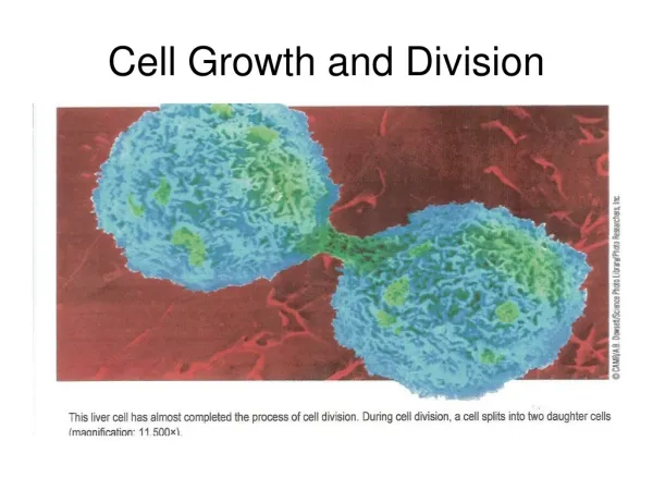
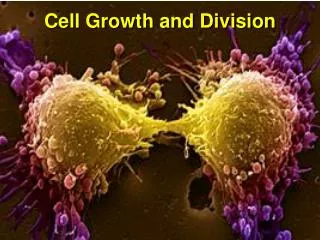
Mitosis (M) • Mitosis includes two processes: mitosis and cytokinesis. • Mitosis is the division of the cell nucleus and its contents. • The nuclear membrane dissolves, duplicated DNA condenses around proteins and separates, and two new nuclei form. • Cytokinesis is the process that divides the cell cytoplasm. • The result is two daughter cells that are genetically identical to the original cell.
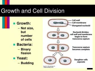
Cells Divide At Different Rates • The rate at which your cells divide is linked to your body’s need for those cells. • The internal lining of your digestive track receives a lot of wear and tear. As a result, cells that line your stomach and intestine are replaced every few days. • Cells that make up the smooth muscle of the intestine, the lungs, kidney, and liver, divide only occasionally in response to injury or death.
G0 • Cells that divide only rarely are thought to enter a stage called G0. • G0 cells are unlikely to divide, although they continue to carry out their normal functions. • Some cells, such as neurons, appear to stay permanently in the G0 stage. • Lymphocytes, a type of white blood cell, may remain in the G0 stage for years until they recognize an invader. • Once the invader binds to a lymphocyte receptor, the lymphocyte goes through rapid cell divisions to help fight infection. Nerve cell communicating via neurotransmitters Lymphocyte
Cell Size Is Limited • Cells have upper and lower size limits. • If cells were too small, they could not contain all the necessary organelles and molecules. • The upper limit of cell size is due to the ratio of cell surface area to volume. • Oxygen, nutrients, and wastes move across the cell membrane, or the surface of a cell. • These materials must be transported in adequate amounts and with adequate speed to keep the inside of the cell functioning. • As a cell increases in size, its volume increases faster than its surface area. • Therefore, a further increase in size could result in a surface area too small for the adequate exchange of materials. A neuron running down a giraffe’s neck to its legs may be several meters long, but it is not shaped like a cube or sphere. It is extremely long and thin. This structure gives the neuron a large surface area with a relatively small increase in volume.
Mitosis and Cytokinesis • Mitosis is an amazing process that efficiently sorts two sets of DNA and divides them between two nuclei.
Chromosomes Condense • DNA is a double-stranded molecule made of four different subunits called nucleotides. • A chromosome is one long continuous thread of DNA that consists of numerous genes along with regulatory information. • Your body cells have 46 chromosomes each. • If stretched out straight and laid end to end, the DNA in just one of your cells would be about 10 feet long. • During interphase, DNA is loosely organized (like spaghetti). • During mitosis, DNA has to be condensed so it can be divided accurately.
Chromosomes Condense • Each of your chromosomes is associated with a group of proteins called histones. • DNA wraps around histones at regular intervals like beads on a string. • When DNA is loosely wrapped (during interphase) it is called chromatin. • As a cell progresses into mitosis, chromatin further condenses, until it forms small thick rods.
Chromosomes Condense • The chromosome now looks like an ‘X’. • One half of the duplicated chromosome is called a chromatid. • Each chromosome is held together in the center by a centromere. • The ends of the DNA molecule form telomeres. • Telomeres are sequences of repeating nucleotides that do not form genes. • They prevent the ends of chromosomes from accidently attaching to each other, and they prevent the loss of genes.
Interphase • Interphase prepares the cell to divide. • It provides critical time for the duplication of organelles and for DNA replication. • By the end of interphase, an individual cell has two full sets of DNA, or chromosomes, and is large enough to divide.
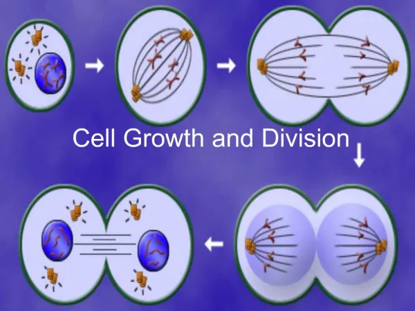
Mitosis • Mitosis divides a cell’s nucleus into two genetically identical nuclei, each with its own single, full set of DNA. • The four main stages of mitosis are: • Prophase • Metaphase • Anaphase • Telophase
Mitosis: Prophase • Chromatin condenses into tightly coiled chromosomes. • Each consists of two identical sister chromatids. • The nuclear envelop breaks down, the nucleolus disappears, and the centrosomes and centrioles begin to migrate to opposite sides of the cell. • Organized microtubules called spindle fibers grow from the centriolesand radiate toward the center of the cell.
Mitosis: Metaphase • The spindle fibers attach to a protein structure on the centromere of each chromosome and align the chromosomes along the cell equator, around the middle of the cell.
Mitosis: Anaphase • Sister chromatids separate from each other. • Spindle fibers begin to shorten, which pulls the sister chromatids away from each other and toward opposite sides of the cell.
Mitosis: Telophase • A complete set of identical chromosomes is positioned at each pole of the cell. • The nuclear membranes start to form, the chromosomes begin to uncoil, and the spindle fibers fall apart.
Cytokinesis • Cytokinesis divides the cell into two cells and completes a full stage of the cell cycle. • In animal cells, the membrane forms a furrow, or trench, that is pulled inward by tiny filaments, like a drawstring. • Gradually, the membrane pinches closed, forming a separate cell around each nucleus. • In plant cells, a cell plate is formed between the two nuclei. • It is made by the golgi apparatus, which supplies the new plasma membrane. Cleavage furrow in animal cell Cell plate in plant cell
Regulation of the Cell Cycle • How does your body regulate all the millions of cells divisions happening in your body?
External Factors • External factors that help regulate the cell cycle include physical and chemical signals. • One physical factor is cell-cell contact. • Once a cell touches another cell, it will stop dividing. • A chemical factor is growth factor. • Growth factors are a broad group of proteins that stimulate cell division. Growth factors bind to receptors that activate specific genes to trigger cell growth. There are many different types of growth factor. • Platelets let out a growth factor that stimulates all types of cells around a wound to begin the healing process. • Erythropoietin stimulate the production of red blood cells. • Growth hormone results in bone growth and affects your protein and fat metabolism.
Internal Factors • Kinases and cyclins are internal factors that regulate cell division. • A kinase is an enzyme that energizes a molecule and forces it to change shape or to become energized. • Kinases that help the cell cycle are activated by cyclins. • Cyclins are a group of proteins that are rapidly made and destroyed at certain points in the cell cycle. • These two factors help cells advance to different stages of the cell cycle.
Apoptosis • Apoptosis is programmed cell death. • This occurs when internal or external signals activate genes that help produce self-destructive enzymes. • Cells in the immune system recognize apoptopic cells and eat them. • For example: in early stages of development, human embryos have webbing between their fingers and toes. Before a baby is born, those cells typically go through apoptosis, and most babies are born with clearly defined, un-webbed fingers and toes.
Uncontrolled Division • Cancer is the common name for a class of diseases characterized by uncontrolled cell division. • It arises when regulation of the cell cycle breaks down. • Cancer cells continue to divide even when they come into contact with other cells. They will also divide in the absence of growth factors. As a result, they divide much more often then healthy cells. Breast cancer cells
Uncontrolled Division • Cancer cells form disorganized clumps called tumors. • Benign tumors are when cancer cells typically remain clustered together. • This means that the tumor may be relatively harmless and can probably be cured by removing it. • Malignant tumors are when cancer cells break away from the main tumor. This is called metastasizing. • The breakaway cells can be carried in the bloodstream or lymph system to other parts of the body where they can form more tumors. • These are very difficult to remove. Benign tumor Malignant tumor
Causes of Uncontrolled Division • Some cancers are caused by cells that have suffered damage to the genes that help make proteins involved in cell-cycle regulation. • Most canter cells carry mutations, or errors, in two types of genes. • One type, oncogenes, accelerate the cell cycle, the other type acts as cell-cycle brakes. • Mutations in these genes can be inherited. • Some mutations can be caused by exposure to radiation (UV light) or chemicals. • Substances known to produce or promote the development of cancer are called carcinogens. • These include tobacco smoke and certain air pollutants. • Other cancers can be caused by viruses. • HPV is known to cause cervical cancer.
Treatments for Cancer • Standard treatment often involves both radiation and chemotherapy. • Radiation therapy is the use of radiation to kill cancer cells and shrink tumors. It works by damaging a cell’s DNA so much that the cell cannot divide. • Radiation is usually localized because it can also hurt healthy cells. • Chemotherapy uses certain drugs, often in combination, to kill actively dividing cells. • Chemotherapy is systemic—drugs travel throughout the entire body. Susan G. Komen was diagnosed with breast cancer at the age of 33. She succumbed to the disease three years later in 1980. Her sister made a promise to increase research and development of a cure for breast cancer, and founded the Susan G. Komen Breast Cancer Foundation in 1982.