Download
1 / 31
310 likes | 475 Views
DNA & Protein Synthesis Chapter 18. The Molecule of Heredity. Seeking the Genetic Material. 1928 Griffith finds that virulent bacteria can transform nonvirulent bacteria into the deadly form. Virulent : able to cause disease

E N D
DNA & Protein SynthesisChapter 18 The Molecule of Heredity
Seeking the Genetic Material • 1928 Griffith finds that virulent bacteria can transform nonvirulent bacteria into the deadly form. Virulent: able to cause disease • 1944 Avery: found DNA was the molecule of heredity, not protein or RNA. • 1952 Hershey and Chase: found that viruses injected DNA into host bacteria. DNA is confirmed as the unit of heredity. Animation & Game Avery, Macleod, McCarthy Hershey and Chase Experiment
Who Else Contributed? • Chargaff:1949 discovered there are always equal amounts of Adenine to Thymine and the same percentage of Cytosine to Guanine. • Rosalind Franklin 1950-53: Photographed DNA with X-rays.(pg 187, Fig 10-4) Found helictical shape (i.e. Helix Shape) while working in the lab of Maurice Wilkins. Chargaff’s Ratios Franklin Click: NOVA News Minute
16.7 The Structure of DNA • Structure was discovered by Watson and Crick in 1953, 1962 receive Nobel Prize. • Double Helix: • Nucleotides: Phosphate, Sugar (deoxyribose) and a Nitrogen base (A,T,C,G) • DNA Backbone: Sugar-Phosphate-Base Connected by Phosphodiester linkage • 5 carbon sugar and phosphate molecule are the same for each nucleotide. Nitrogen base changes.
Deoxyribonucleic Acid (DNA Continued) • 2 strands one strand runs 5’ to 3’ the other 3’ to 5’ (upside down) • Where does the 5’ and 3’ come from? Read DNA From Here Synthesize from 5’ to 3’
Nitrogen Bases: 16.8 • Adenine double bonds to Thymine • Cytosine triple bonds with Guanine • GCAT Bases are held together by weak hydrogen bonds. • Purines (A&G) vs. Pyrimidines (C,T,U) • A mistake here is a Point Mutation. • Can you tell me the complementary strand for : AATCGCGA? ______________
Eukaryotic DNA Replication • Overview Cinema: The Forest from the Trees • The DNA Unzips, and each strand serves as a template for the formation of a new strand composed of complementary nucleotides G-C, A-T HHMI Replication
Replication: Entering the Forest • DNA Replication begins at special Sites called Origins of Replication • Where the two strands of DNA split is called replication bubbles (thousands) • Why do you need thousands of these bubbles? (Hint: The DNA molecule has 3 billion base pairs)
DNA Replication 16.12 • Double helix unwinds using DNA Helicase. • DNA Helicase breaks the hydrogen bonds. • Where the DNA breaks apart is called the replication fork. DNA polymerase (another enzyme) adds nucleotides at the 5’ to 3’ end towards the replication fork. • FYI: Humans= 50 nuclotides/sec
Meselson & Stahl Experiment DNA Replication Explained
What about the 3’ to 5’ end? • Need RNA Primer. (RNA and the enzyme primase). • Lagging strand- away from the replication fork replicates using Okazaki fragments. • Each 100-2000 nucleotide Okazaki fragment is joined by DNA ligase. • Fig 16.15 & 16.16
Checking for errors • DNA Polymerase also proof reads the strands- Mismatch Repair • A mistake in nucleotide pairing is a Mutation • Multiple replication forks happen all at once so that the process is speedy. DNA Review Flashcards
How do we keep the replication process clean? • Single-stranded binding proteins scaffold the two DNA strands apart. • Topoisomerase cuts DNA that is wound too tightly. • Telomeres, made by the enzyme telomerase protect against gene loss at the end of the chromosome. Fig 16.19 • Telomeres repeating sequences of TTAGGG (thousands of times) • Telomeres shorten with each cell division. Aging anyone?
How do bacteria replicate their DNA? Fig. 16.2 The Technical Details The Non-Technical Details The Technical Details
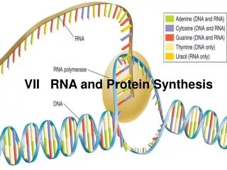
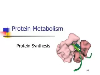
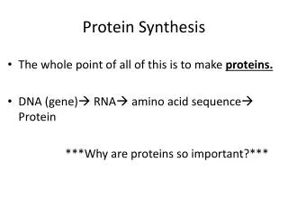
What is RNA and how is it useful? • RNA= Ribonucleic Acid • Single stranded. Ribose sugar. • Transcribes DNA and Translates it into proteins. • Proteins are organic compounds that have specific jobs in the cell. (Ex. Enzymes) PBS Video RNAi
What are the 3 types of RNA? • mRNA= Messenger RNA • Transcribes or rewrites DNA’s message as mRNA, mRNA carries message to ribosome • rRNA = Ribosomal RNA. Fig. 17.16 • Creates the ribosomes on the rough ER and cytoplasm where proteins are made. • tRNA = Transfer RNA. Fig. 17.14 & 17.15 • Transfers amino acids to the ribosomes and translates the mRNA into protein. (Called Translation because the message changes from nucleic acid to protein, a different organic compound)
Transcription and TranslationCinema Fig. 17.3 • Don’t relax too much • Pencils out? Notes ready? Lets work! Overview Movie Start Here HHMi Transcription More Detail
AUG (start) methionine UAA UGA UAG (stop)
What is Wobble? • A relaxation of the base pairing rule. • Example: UCU, UCC, UCA, UCG = serine • Why is their Wobble room? • Allows for point mutations not to cause an incorrect protein to be made. • Since there are only 20 Amino Acids and 64 codons to code for them we can have repeats.
What are the three stages of transcription? • Initiation: RNA polymerase binds to the DNA promoter. Fig 17.8 • TATA box is the area that transcription factors recognize within the promoter. • Creates a transcription initiation complex (transcription factors & RNA polymerase bond on promoter) • Elongation: DNA template continues to grow as RNA polymerase separates DNA strand. • Transcribed part: Transcription unit (made of codons) (1 gene transcribed many times in caravan fashion) • Termination: mRNA is cut free from DNA template
How do Eukaryotes control Gene expression? • Transcription creates Pre-mRNA. Fig 17.10 • Pre-mRNA includes Introns and Exons • Introns= Fillers (intervening sequences) • Exons= Code for proteins (expressed sequences) • mRNA is just the Exons Eukaryotic Transcription
How else is mRNA processed for translation? • snRNP’s (“snurps”) & splicesomes remove introns, creating the final mRNA sequence= Exons only. Fig. 17.11 • What stops the mRNA from degrading by hydrolytic enzymes & binds it to the ribosome? • 5’ cap: modified guanine nucleotide is added to the 5’ end. • A poly tail: 30-200 adenine nucleotides on the 3’ end. (Also helps release mRNA into the cytoplasm.
What do enhancers do? Fig 18.9 • Enhancer’s causes the gene it is enhancing to be expressed. Transcription occurs in eukaryotes when an enhancer site is activated.
Fig. 18-9-3 Promoter Activators Gene DNA Distal control element Enhancer TATA box General transcription factors DNA-bending protein Group of mediator proteins HHMI Transcription Basic RNA polymerase II RNA polymerase II Transcription initiation complex RNA synthesis
Fig. 18-8-3 Putting it all together Poly-A signal sequence Enhancer (distal control elements) Proximal control elements Termination region Exon Intron Exon Intron Exon DNA Upstream Downstream Promoter Transcription Exon Intron Exon Intron Exon Primary RNA transcript Cleaved 3 end of primary transcript 5 RNA processing Intron RNA Poly-A signal Coding segment mRNA 3 Start codon Stop codon Poly-A tail 3 UTR 5 Cap 5 UTR
And now Translation Fig. 17.17-17.19 • Translation: mRNA codons code for an Amino acid sequence. • tRNA attaches to the ribosomal rRNA with anticodon. GTP recycles tRNa with enzyme. • Ribosome: protein and rRNA created in the nucleolus. • Bacterial & Eukaryotic ribosomes differ, this allows Tetracycline and Streptomycin to stop protein production in bacterial ribosomes. • Small and large subunits for attachment to mRNA. • Three binding sites for tRNA • P site: Holds the growing polypeptide chain • A site: Holds the newest tRNA • E site: Exit site
What are the three stages of translation? • Initiation: mRNA’s AUG codon (methionine) attaches to the ribosome. tRNA attaches. • Elongation: forms the polypeptide chain as tRNA’s continue to attach. • Termination: mRNA reaches a stop codon. Release factor hydrolyzes the bond. mRNA is degraded. Polypeptide is freed from ribosome. • After this the polypeptide will fold or pleat (secondary structure), Chaperonine will complete the tertiary structure. How Proteins fold Fold it, the Game!!!
What about when a mistake occurs? • They can effect the species gene pool. • Point mutation= single base-pair substitution. (sickle cell) • Insertion or deletion= frameshift mutations. The Dog -> HeD og • Missence Mutation= If a codon within a gene turns into a stop codon translation is altered.
Gel Electrophoresis • How does this link to Gel Electrophoresis? • Below is a reminder of what Electrophoresis is and how it works. Gel Electrophoresis