Download
1 / 11
110 likes | 225 Views
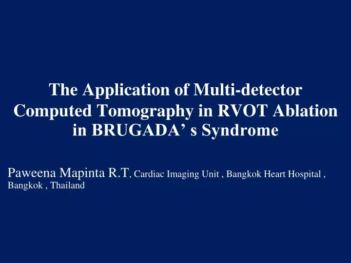
The Application of Multi-detector Computed Tomography in RVOT Ablation in BRUGADA’ s Syndrome. Paweena Mapinta R.T , Cardiac Imaging Unit , Bangkok Heart Hospital , Bangkok , Thailand. Background.

E N D
The Application of Multi-detector Computed Tomography in RVOT Ablation in BRUGADA’ s Syndrome Paweena Mapinta R.T, Cardiac Imaging Unit , Bangkok Heart Hospital , Bangkok , Thailand
Background • Radiofrequency ablation (RFA) is the effective treatment of the right ventricular outflow tract (RVOT) tachycardia and Brugada’s syndrome . MDCT has role in RFA planning by demonstrating the image of the right ventricular outflow tract (RVOT) area which is the target for ablation • In adult patient , coronary artery disease is always the important condition which must be assessed before ablation operation. MDCT scanning with contrast for RVOT and for coronary artery evaluation may be requested. Although double CT scanning can provide all needed information but radiation and iodinated contrast exposure dose are increased
Research Question The newly modified technique(single scan with iodinated contrast dose adjusting) which reduces radiation and contrast dose exposure can be used effectively in demonstrating the RVOT and coronary artery comparable to the conventional technique?
Objective • To prove that the newly modified technique which reduces theradiation and contrast doses can be used effectivelycomparable to the conventional technique indemonstrating the RVOT and coronary artery anatomicalstructure before ablation operation without degradation of theimage quality
Population and method • 14 cases of the Brugada’s syndrome patients who underwent MDCT scan were recruited since the year of 2008 to 2011,12 patients were male • The mean age of the patient was of 47.5 years old • 12 patients were scanned by 256- slice MDCT,2 patients were scanned by 64-slice MDCT. The newly modified technique was used for all patients • The quality of the images obtained by the modified technique were assessed in terms of readable and mapping qualification by ROI of the interest area > 300 HU
Results • The optimum added volume of contrast using of MDCT with the newly modified technique for RVOT and coronary artery scanning in one procedure is 15% of calculated contrast volume • MDCT using newly modified technique provided good quality images of coronary artery and RVOT in terms of ROI>=300 for 64.28% and readable for 100%
Conclusion • The newly modified techniquewhichreducesthe radiationand contrast doses can be used successfully in demonstrating RVOT and coronaryartery before ablation operation without degradation of the imagequality
Conventional technique image RVOT RVOT Aorta Aorta
Modified technique image RVOT RVOT Aorta Aorta