Download
1 / 31
310 likes | 430 Views
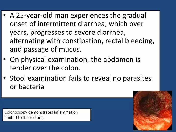
A 25-year-old man experiences the gradual onset of intermittent diarrhea, which over years, progresses to severe diarrhea, alternating with constipation, rectal bleeding, and passage of mucus. On physical examination, the abdomen is tender over the colon.

E N D
A 25-year-old man experiences the gradual onset of intermittent diarrhea, which over years, progresses to severe diarrhea, alternating with constipation, rectal bleeding, and passage of mucus. • On physical examination, the abdomen is tender over the colon. • Stool examination fails to reveal no parasites or bacteria Colonoscopy demonstrates inflammation limited to the rectum,
Biopsy Active chronic colitis. Normal
What histologic feature is seen in Crohn disease that is not seen in ulcerative colitis? What are the complications of ulcerative colitis? Granulomas and transmural inflammation in the resected specimen. The most serious complication is the development of carcinoma. The cancers are preceded by dysplasia, which tends to arise in multiple sites. The risk of cancer is highest in patients with pancolitis of ten or more years duration, in whom it is 20 to 30 fold higher than in a control population. Other life-threatening complications include severe diarrhea and electrolyte disturbances, severe colonic dilation (toxic megacolon) with potential for perforation and peritonitis, and massive hemorrhage.
A 25-year-old man presents to a rheumatologist with complaints of joint pain involving the large joints of the legs. On questioning, the patient indicates that exacerbations in the joint pain are frequently accompanied by diarrhea. Which of the following gastrointestinal diseases is most likely to be implicated as the cause of the patient's joint problems? • A. Amebic colitis • B. Chronic appendicitis • C. Diverticulosis • D. Pseudomembranous colitis • E. Ulcerative colitis
Mucosal ulceration is seen in both Crohn's disease and ulcerative colitis. The ulcers of Crohn's disease are generally described as linear fissures, following the longitudinal axis of the intestine. Ulcerative colitis typically produces broad, extensive areas of ulceration. Pseudopolyps are most commonly associated with ulcerative colitis, and represent the islands of spared mucosa between the broad ulcerations. Rectal involvement in inflammatory bowel disease is more typical of ulcerative colitis than of Crohn's disease. Whereas ulcerative colitis is a "pancolitis," that is usually most severe in the rectum and right colon, Crohn's disease is usually a disease of the small intestine, and may involve the small intestine alone (40%) or both the small intestine and colon (30%). • Crohn's disease is frequently associated with "skip lesions,“ discontinuous areas of active disease in the colon and small intestine with intervening segments that appear normal. This is in marked contrast to ulcerative colitis, which most commonly shows continuous mucosal involvement. Both ulcerative colitis and Crohn's disease can show mucosal atrophy. . Chronic mucosal inflammation produces glandular atrophy, and a loss of mucosal folding.
A patient has had years of intermittent diarrhea and abdominal pain, but has never consulted a physician. Eventually, he begins to pass fecal material in his urine and he seeks medical attention. Which of the following diseases is most likely to cause this complication? • A. Celiac disease • B. Crohn's disease • C. Diverticulitis • D. Ulcerative colitis • E. Whipple's disease
The correct answer is B. Passing fecal material in urine strongly suggests the possibility of a fistula between the bowel and bladder. Of the diseases listed, only Crohn's disease (a type of inflammatory bowel disease) commonly produces fistulas. Fistulas are produced in Crohn's because the disease affects the entire thickness of the bowel wall, rather than being restricted to the mucosa (e.g., ulcerative colitis).
35 y/o male , known case of inflammatory bowel disease, presented in ER with severe colicky abdominal pain. Barium enema study show features of intestinal obstruction • He was taken to OR , and Excision of terminal ileum and proximal colon was performed.
Thickened ileum Right colon Normal ileum The specimen is a section of normal ileum, thickened ileum, and right colon. The intestinal wall is thick, the result of edema, inflammation, fibrosis, and hypertrophy of the muscularispropria. Linear ulcers are typically present in the diseased segment of bowel. In diseased bowel segments, the serosa is thickened and fibrotic, and often the mesenteric fat wraps around the bowel surface (creeping fat).
crohn’s disease crypt distortion Noncaseatinggranulomas Submucosal edema, fibrosis, and lymphoid aggregates
What are the complications of Crohn disease? Fissures in the mucosa can extend through the wall and form sinus tracts, resulting in fistula formation to other loops of bowel, urinary bladder or vagina; there may be localized peritonitis and abdominal abscesses; Fibrosis of the gut wall may lead to strictures and obstruction. Extensive involvement of the small bowel may cause marked loss of albumin (protein-losing enteropathy) or malabsorption.
Are any other organs affected in Crohn disease? In ulcerative colitis? Both Crohn disease and ulcerative colitis are systemic diseases, associated with varied extraintestinal manifestations of immunologic origin. These include polyarthritis, sacroiliitis, ankylosingspondylitis, uveitis, sclerosingcholangitis, erythemanodosum, and clubbing of the fingertips.
A 39-year-old male presents with bloody diarrhea. Multiple stool examinations fail to reveal any ova or parasites. A colonoscopy reveals the rectum and sigmoid portions of the colon to be unremarkable. A biopsy from the terminal ileum reveals numerous acute and chronic inflammatory cells within the lamina propria. Worsening of the patient’s symptoms results in emergency resection of the distal small intestines. Gross examination of this resected bowel reveals deep, long mucosal fissures extending deep into the muscle wall. Several transmural fistulas are also found. What is the best diagnosis for this patient? a. Ulcerative colitis b. Lymphocytic colitis c. Infectious colitis d. Eosinophilic colitis e. Crohn’s disease
CD is classically described as being a granulomatous disease, but granulomas are present in only 25 to 75% of cases. Therefore, the absence of granulomas does not rule out the diagnosis of CD. CD may involve any portion of the gastrointestinal tract and is characterized by focal (segmental) involvement with “skip lesions.” Involvement of the intestines by CD is typically transmural inflammation, which leads to the formation of fistulas and sinuses. The deep inflammation produces deep longitudinal, serpiginous ulcers, which impart a “cobblestone” appearance to the mucosal surface of the colon. Additionally in Crohn’s disease, the mesenteric fat wraps around the bowel surface, producing what is called “creeping fat,” and the thickened wall narrows the lumen, producing a characteristic “string sign” on x-ray. This narrowing of the colon, which may produce intestinal obstruction, is grossly described as a “lead pipe” or “garden hose” colon. • In contrast to CD, UC affects only the colon, and the disease involvement is continuous. The rectum is involved in all cases, and the inflammation extends proximally. Since UC involves the mucosa and submucosa, but not the wall, fistula formation and wall thickening are absent (but toxic megacolon may occur). Grossly, the mucosa displays diffuse hyperemia with numerous superficial ulcerations. The regenerating, nonulcerated mucosa appears as “pseudopolyps.
Inflammatory bowel diseases Crohn's disease Ulcerative Colitis • Colon only • Diffuse involvement of mucosa • Superficial ulcers • Any part of the GIT • Skip areas of normal mucosa • Mucosal inflammation only • Fistula formation • Transmural inflammation • Granulomas • Deep ulcers ( fissure ) • Dysplasia is common • Carcinoma is more common (10%)
1. What is the most likely diagnosis with this presentation? A 68-year-old woman presents with intermittent constipation, weight loss, and a swollen abdomen. She has had two previous polypectomies: one showed a tubular adenoma and the other was a tubulovillous adenoma. Double-contrast barium enema shows an irregular stricture 4 cm long in the ascending colon. A tumor is diagnosed and surgery is advised. The tumor is resected and is found to have invaded through the thickness of the bowel wall, but is completely excised. Three of 15 lymph nodes identified contained metastatic tumor. 1. The most likely diagnosis is colorectal adenocarcinoma. 2. What stage is this tumor and what is the prognosis? 2. This is a Dukes’ C carcinoma (T3, N1, MX
3. What is the association between adenoma and carcinoma? There is much evidence to suggest that most carcinomas of the colon arise in pre-existing adenomas (adenoma–carcinoma sequence). Patients with familial adenomatouspolyposis (FAP) have a very high risk of developing colorectal carcinomas. 4. Where is the metastatic spread most likely? 4. Colorectal carcinomas metastasize mainly to regional lymph nodes and liver, less commonly developing other systemic metastases such as brain, bone and lung.
52 y/o female presented with fatigue and weakness. She experienced 6 kg wt loss in a 6 six months CBC .....Hg 7.5 g/dl, hematocrit 26 % Serum ferritin 8 ng/dl Iron deficiency anaimia Rx oral iron treatment
Stool and urine analysis……blood in the stool Colonoscopy …….ascending colon 6 cm mass Biopsy ……..adenocarcinoma
Colonic polyposis Multiple osteoma Central nervous system tumors Fibromatosis 100% risk of carcinoma Cutaneous cysts Familial polyposis coli Gardner’s syndrome Turcot’s syndrome.
Polyps • Non-neoplastic polyps 90% • Neoplastic polyps 10%
Polyps • Non-neoplastic polyps 90% • Hyperplastic polyps • Hamartomatous polyps • Inflammatory polyps • Lymphoid polyps • Neoplastic polyps 10% • Adenoma
Polyps • Non-neoplastic polyps 90% • Hyperplastic polyps • Hamartomatous polyps (Juvenile & Peutz-Jeghers polyps) • Inflammatory polyps • Lymphoid polyps • Neoplastic polyps 10% • Adenoma
A 22-year-old woman has had recurrent episodes of diarrhea, crampy abdominal pain, and slight fever over the last 2 years. Other symptoms have included mild joint pain and sometimes red skin lesions. On at least one occasion, her stool has been iron-positive, indicating the presence of occult blood. Colonoscopy reveals several sharply delineated areas with thickening of the bowel wall and mucosal ulceration. Areas adjacent to these lesions appear normal. Biopsies of the affected areas show full-thickness inflammation of the bowel wall and several noncaseatinggranulomas. • · What is the most likely diagnosis? • · What are the common complications of this disease?
Most likely diagnosis:Crohn disease. • · Common complications of this disease:Malabsorption and malnutrition, fibrous strictures of the intestine, and fistulae to other organs, such as from bowel to skin or bowel to bladder.
A 26-year-old man presents with intermittent crampy abdominal pain, diarrhea without noticeable blood, and weight loss of 15 lb over 10 months. The bowel symptoms, including the diarrhea, wake him from sleep; he resumed smoking cigarettes a year ago. His older brother has had similar symptoms but has not yet been evaluated. Stool leukocytes are present. Results of examination with sigmoidoscopy are normal. Which of the following is the most likely diagnosis for this patient? • ❑ A. Irritable bowel syndrome • ❑ B. Acute appendicitis • ❑ C. Crohn disease • ❑ D. Ulcerative colitis • ❑ E. Colon cancer
Which of the following statements regarding the relationship between colon cancer and polyps is false? ❑ A. Most colorectal cancers arise from preexisting adenomas ❑ B. Adenomatous polyps, as well as juvenile polyps, hamartomas, and inflammatory polyps, progress to colorectal carcinoma ❑ C. Larger polyps, especially those larger than 1 cm, are more likely to contain invasive carcinoma ❑ D. On the basis of histology, villous polyps are more likely to contain invasive carcinoma than are tubular polyps