Download

1 / 61
670 likes | 910 Views
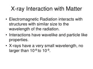
X-ray Interaction with Matter. Electromagnetic Radiation interacts with structures with similar size to the wavelength of the radiation. Interactions have wavelike and particle like properties. X-rays have a very small wavelength, no larger than 10 -8 to 10 -9. X-ray Interaction with Matter.

E N D
X-ray Interaction with Matter • Electromagnetic Radiation interacts with structures with similar size to the wavelength of the radiation. • Interactions have wavelike and particle like properties. • X-rays have a very small wavelength, no larger than 10-8 to 10-9.
X-ray Interaction with Matter • The higher the energy of the x-ray, the shorter the wavelength. • Low energy x-rays interact with whole atoms. • Moderate energy x-rays interact with electrons. • High energy x-rays interact with the nuclei.
Five forms of x-ray Interactions • Classical or Coherent Scattering • Compton Effect • Photoelectric Effect • Pair production • Photodisintegration
Two Forms of X-ray Interactions Important to Diagnostic X-ray • Compton Effect • Photoelectric Effect
Classical or Coherent Scattering • Low energy x-rays of about 10 keV interact in this manner. • Incident photon interacts with the atom. • There is a change in direction.
Classical or Thompson Scattering • There is no loss of energy and no ionization. • Photon scattered forward. • Because these are low energy x-rays, they are of little importance.
Classical Scattering • At 70 kVp only a few percent of the x-rays undergo this form of scattering. • Classic Scatter may contribute to the graying of the image called film fog.
Compton Effect • Moderate energy x-ray photon through out the diagnostic x-ray range can interact with outer shell electron. • This interaction not only changes the direction but
Compton Effect • reduced its energy and ionizes the atom as well. The outer shell electron is ejected. This is called Compton Effect or Compton Scattering.
Compton Scattering • The x-ray continues in an altered direction with decreased energy. • The energy of the Compton-scattered x-ray is equal to the difference between the energy of the incident x-ray and the energy imparted to the electron.
Compton Scattering • The energy imparted to the electron is equal to its binding energy plus the kinetic with which it leaves the atom. • During Compton-scattering most of the energy is divided between the scattered photon and the secondary electron. • The Secondary Electron is called a Compton Electron.
Compton Scattering • The scattered photon and secondary electron will retain most of its energy so it can interact many times before it losing all of it’s energy.
Compton Effect • The scattered photon will ultimately be absorbed photoelectrically. • The secondary electron will drop into a hole in the outer shell of an atom created by an ionizing event. • Compton-scattered photons can be deflected in any direction.
Compton Effect • A zero angle deflection will result in no energy loss. • As the angle approaches 180 degrees, more energy is transferred to the secondary electron. • Even at 180 degrees, 66% of the energy is retained.
Compton Effect • Photons scattered back towards the incident x-ray beam are called Backscatter Radiation. • While important in radiation therapy, backscatter in diagnostic x-ray is sometimes responsible for the hinges on the back of the the cassette to be seen on the x-ray film
Compton Effect • The probability of Compton Effect is about the same for soft tissue or bone. • This decreases with increasing photon energies. • Compton scatter decreases with increased kVp.
Most likely to occur As x-ray energy increases With outer-shell electrons With loosely bound electrons. Increased penetration through tissue w/o interaction. Increased Compton relative to photoelectric scatter. Reduced total Compton scattering. Features of Compton Scattering
As atomic number of the absorber increases As mass density of absorber increases No effect on Compton Scatter Proportional increase in Compton Scatter. Features of Compton Scatter
Photoelectric Effect • X-rays in the diagnostic range can undergo ionizing interactions with inner shell electron of the target atom. • It is not scattered but totally absorbed.
Photoelectric Effect • The Photoelectric Effect is a photon absorption interaction.
Photoelectric Effect • The electron removed from the target atoms is called a photoelectron. • The photoelectron escapes with kinetic energy equal to the difference between the energy of the incident x-ray and the binding energy of the electron.
Photoelectric Effect • Low anatomic number target atoms such as soft tissue have low binding energies. • Therefore the photoelectric electron is released with kinetic energy nearly equal to the incident x-ray. • Higher atomic number target atoms will have higher binding energies.
Photoelectric Effect • Therefore the kinetic energy of the photoelectron will be proportionally lower. • Characteristic x-rays are produced following a photoelectric interaction to those produced in the x-ray tube. • These characteristic x-rays are also secondary radiation and acts like scatter.
Photoelectric Effect • The probability of a photoelectric interaction is a function of the photon energy and the atomic number of the target atom. • A photoelectric interaction can not occur unless the incident x-ray has energy equal to or greater than the electron binding energy.
Photoelectric Effect • The probability of photoelectric interaction is inversely proportional to the third power of the photon energy. • The probability of photoelectric interaction is directly proportional to the third power of the atomic number of the absorbing material
Human Tissue Muscle Fat Bone Lung Other Material Air Concrete Lead Effective Atomic # 7.4 6.3 13.8 7.4 7.6 17 82 Effective Atomic Numbers
Photoelectric Effect • A probability of interaction to the third power changes rapidly. • For the photoelectric effect this means that a small variation in atomic number or x-ray energy results in a large changes in chance of an interaction. • This is unlike Compton interactions.
Most likely to occur With inner-shell electrons With tightly bound electrons. When the x-ray energy is greater than the electron-binding energy. Features of the Photoelectric Effect
As the x-ray energy increases Increased penetration through tissue without interaction. Less photoelectric effect relative to Compton effect. Reduced absolute absorption. Features of the Photoelectric Effect
As the atomic number of the absorber increases As mass density of the absorber increases Increases proportionally the cube of the Z. Proportional increase in photoelectric effect. Features of the Photoelectric Effect
Pair Production • If the incident x-ray has sufficient energy, it may escape the electron cloud and come close enough to the nucleus to come under the influence of the strong electrostatic field of the nucleus.
Pair Production • The interaction with the nucleus strong electrostatic field causes the photon to disappear and in its place appear two electrons.
Pair Production • One is positively charged and called a positron while the other remains negatively charged. This is called Pair Production.
Pair Production • It take a photon with 1.02 MeV to undergo Pair Production. • Therefore it is not important to diagnostic x-ray.
Photodisintegration • High energy x-ray photons with energies above 10 MeV can escape interaction with both the electrons and nucleus electrostatic fields.
Photodisintegration • It is absorbed into the nucleus that excites the nucleus resulting in the release of a nucleon or other nuclear material. This is referred to as:
Photodisintegration • Photodisintegration. Like pair production, the high energy needed to cause this makes it unimportant to diagnostic radiography.
Differential Absorption • Only Compton and Photoelectric Effects are important interactions that the x-ray may have with matter in the diagnostic spectrum. • More important than the x-rays resulting from these effects are a third type, those transmitted through the body without interacting.
Differential Absorption • Those that make it through the body contribute to the radiograph. • It should be clear than Compton Scatter X-rays contribute no useful information. • The film does not recognize the scattered x-rays as representing an interaction of the straight line from the target.
Differential Absorption • These scattered x-rays result in film fog, a generalized dulling of the image on the radiograph by film densities not representing diagnostic information. • To reduce this type of fog, we use techniques and apparatus to reduce the amount of scatter reaching the film.
Differential Absorption • X-rays that undergo photoelectric interaction provide diagnostic information to the image receptor. • Since they do not reach the film, these x-rays are representative of anatomic structures with high x-ray absorption characteristics. These structures are said to
Differential Absorption • Be Radiopaque. • The other x-rays that penetrate the body and are transmitted without interaction are said to be Radiolucent. Radiolucent matter appears as high density or dark areas on the radiograph.
Radiopaque Radiolucent Appears Bright Appears Dark Differential Absorption
Differential Absorption • The radiographic image is the result of the difference between those x-rays absorbed photoelectrically and those not absorbed at all. • This characteristic is called differential absorption.
Differential Absorption • Except at very low kVp, most x-rays that interact do so by the Compton effect; this is one reason why radiographs are not as sharp as photographs. • As a rule of thumb, less than 5% of x-rays incident on the patient reaches the film and less than one half of these interact with the film.
Differential Absorption • The radiographic image results from less than 1% of the x-rays emitted from the tube. • Therefore careful control of the x-ray beam is necessary to produce high quality radiographs!
Differential Absorption • Differential Absorption increases as the kVp is lowered but lowered kVp results in a higher patient radiation exposure. • A compromise is needed for each examination.
Differential Absorption • Notice how much of the x-rays are absorbed photoelectrically in bone compared to the soft tissue.
Differential Absorption • The photoelectric absorption of bone is about 7 times greater than in soft tissue regardless of the energy.
Differential Absorption • As kVp is increased fewer interaction occur so more x-rays are transmitted without interaction. • Compton Scatter is independent of the atomic number of the absorbing material and is inversely proportional to the x-ray energy.