Download
1 / 10
100 likes | 268 Views
Structure of repressor protein and how the structure can help understand the binding to various operators. . By Supriya Pokhrel . Lysogenic cycle. Phages reproduce either through a lytic cycle or lysogenic cycle.

E N D
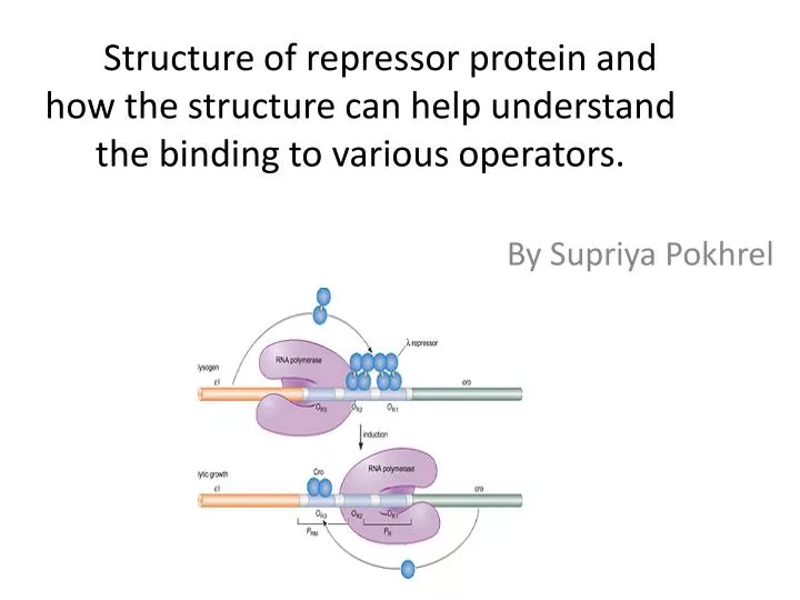
Structure of repressor protein and how the structure can help understand the binding to various operators. By Supriya Pokhrel
Lysogenic cycle • Phages reproduce either through a lytic cycle or lysogenic cycle. • During a lysogenic cycle, phage DNA is integrated into the host chromosome allowing the host to replicate normally. Phages remain dormant until environmental factors compel them to enter the lytic life cycle. • There are some proteins that play an important role in determining mode of replication. Repressor proteins are such type of proteins that control the lysogenic life cycle of various bacteriophages by binding to operators.
Repressor protein and its role in lysogenic phages • Repressor is a product of CI gene. • Repressor binds to operators and enhance repression of promoters to maintain lysogenic cycle. • It was found that L1 and lambda repressors had two domains: C-terminal domain and N-terminal domain (Ganguly et al.). • Repressor protein of lambda phage have been studied and compared to many other lysogenic phages.
C-terminal domain and N-terminal domain • C-terminal domain: Carboxyl terminus is the end of the amino acid chain. It consists of a carboxyl group –COOH. • N-terminal domain: Amino-terminus group refers to the start of the protein and it consists of a free -NH2 group. • During translation, protein starts translation from the N-terminal domain to C-terminal domain.
What is the structure of repressor protein and how can it help understand the binding to operators?
Experimental Method • Lambda repressor consisted of C-terminal domain and N-terminal domain. • These domains were detected using proteolysis of His-CI; two digestive enzymes chymotrypsin and trypsin were used separately(Ganguly et al.)
In lysogenic phages like lambda, a stable lysogen is maintained by the binding of CI to operators (OL and OR). Due to CI’s asymmetric nature, it binds to O64 strongly than OL. This conclusion was based on an experiment using gel shift assays. (L5 phage) A mixture of 20ul buffer A contained repressor and operator DNA was labeled then incubated at 25°C for about 20 minutes. CI had a higher affinity to O64 operator because its distance was shorter (140 nm) than that of OL (370). This proved that operator 64 binds strongly to CI repressor than O left
Results • As a result of proteolysis experiment, CI structure posses both C-terminal domain and N-terminal domain with amino acid residues. • CI had a higher affinity to O64 operator because its distance was shorter (140 nm) than that of OL (370). This proved that operator 64 binds strongly to CI repressor than O left. • The C-terminal domain mediates the interactions of two repressor dimers to operator sites, O64 and OL. The N-terminal domain mediates the interaction with RNA polymerase (lysogenic promoter for repressor maintenance) and thus, activating the CI gene. (Bell et al.) • Therefore, above reasons are why the structure of repressor protein (CI, containing CTD and NTD) is important and how that helps during the binding to operators.
References Mazumder, Abishek, and SumitaBandyopadhyay. "A Genetic Network That Balances Two Outcomes Utilizes Asymmetric Recognition of Operator Sites.” Biophysical Journal Elsevier Inc, Vol 102, issue 107, 4 April 2012, Pages 1580–1589. Bell, Charles E. “Crystal Structure of the λ Repressor C-Terminal Domain Provides a Model for Cooperative Operator Binding.” Cell Press, June 23 2000. Volume 101, 801-811. Ganguly, Tribid and Bandhu, Amitava. “Repressor of temperate mycobacteriophage L1 harbors a stable C-terminal domain and binds to different operator DNAs with variable affinity.” Virology Journal 2007, 4:64.