Download
1 / 118
1.27k likes | 1.35k Views
Diseases of the Pancreas. Victor Politi, M.D., Medical Director SVCMC, School of Allied Health Professions, Physician Assistant Program. Pancreas anatomy. The pancreas is an elongated, tapered organ located across the back of the abdomen, behind the stomach.

E N D
Diseases of the Pancreas Victor Politi, M.D., Medical Director SVCMC, School of Allied Health Professions, Physician Assistant Program
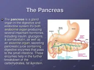
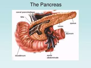
Pancreas anatomy • The pancreas is an elongated, tapered organ located across the back of the abdomen, behind the stomach. • The right side of the organ (called the head) is the widest part of the organ and lies in the curve of the duodenum (the first section of the small intestine). • The tapered left side extends slightly upward (called the body of the pancreas) and ends near the spleen (called the tail).
Pancreas anatomy • The pancreas is made up of two types of tissue: • exocrine tissue • The exocrine tissue secretes digestive enzymes. These are secreted into a network of ducts that join the main pancreatic duct, which runs the length of the pancreas. • endocrine tissue • The endocrine tissue, which consists of the islets of Langerhans, secretes hormones into the bloodstream.
Accessory duct of Santorini • Duct of Wirsung
Pancreas anatomy • The pancreas has digestive and hormonal functions: • The enzymes secreted by the exocrine tissue in the pancreas help break down carbohydrates, fats, and proteins in the duodenum. • These enzymes travel down the pancreatic duct into the bile duct in an inactive form. • When they enter the duodenum, they are activated. • The exocrine tissue also secretes bicarbonate to neutralize stomach acid in the duodenum.
Pancreas anatomy • The hormones secreted by the endocrine tissue in the pancreas are insulin, glucagon (which regulate the level of glucose in the blood), somatostatin (which prevents the release of the other two hormones), and many others.
What is Pancreatitis? • Pancreatitis is an inflammatory process in which pancreatic enzymes autodigest the gland
Normally, digestive enzymes do not become active until they reach the small intestine, where they begin digesting food. • But if these enzymes become active inside the pancreas, they start "digesting" the pancreas itself
The gland can sometimes heal without any impairment of function or any morphologic changes. • This process is known as acute pancreatitis. • It can recur intermittently, contributing to the functional and morphologic loss of the gland. • Recurrent attacks are referred to as chronic pancreatitis.
Acute pancreatitis occurs suddenly and lasts for a short period of time and usually resolves. • Chronic pancreatitis does not resolve itself and results in a slow destruction of the pancreas.
Either form can cause serious complications. • In severe cases, bleeding, tissue damage, and infection may occur. • Pseudocysts, accumulations of fluid and tissue debris, may also develop. • Enzymes and toxins may enter the bloodstream, injuring the heart, lungs, and kidneys, or other organs.
Acute edematous pancreatitis • Since the pancreas is located in the retroperitoneal space with no capsule -inflammation can spread easily. • In acute pancreatitis, parenchymal edema and peripancreatic fat necrosis occur first. • This process is known as acute edematous pancreatitis
Necrotizing pancreatitis • When necrosis involves the parenchyma, accompanied by hemorrhage and dysfunction of the gland, the inflammation evolves into hemorrhagic or necrotizing pancreatitis
Necrotizing pancreatitis • Pseudocysts and pancreatic abscesses can result from necrotizing pancreatitis because of enzymes being walled off by granulation tissue (ie, pseudocyst formation) or bacterial seeding of pancreatic or peripancreatic tissue (ie, pancreatic abscess formation). • An ultrasound or, preferably, a CT scan can be used detect both.
The inflammatory process can cause systemic effects because of the presence of cytokines, such as bradykinins and phospholipase A. • These cytokines may cause vasodilation, increase in vascular permeability, pain, and leukocyte accumulation in the vessel walls. • Fat necrosis may cause hypocalcemia. • Pancreatic B cell injury may lead to hyperglycemia.
Mortality/Morbidity • Although acute pancreatitis should be noted, chronic pancreatitis has a more severe presentation as episodes recur. • Acute respiratory distress syndrome (ARDS), acute renal failure, cardiac depression, hemorrhage, and hypotensive shock all may be systemic manifestations of acute pancreatitis in its most severe form.
Acute Pancreatitis • Some people have more than one attack and recover completely after each, but acute pancreatitis can be a severe, life-threatening illness with many complications.
Acute Pancreatitis • About 80,000 cases occur in the United States each year; some 20 percent of them are severe. • Acute pancreatitis occurs more often in men than women.
The risk for African American persons aged 35-64 years is 10 times higher than for any other group. • African American persons are at higher risk than white persons in that same age group
History • The main presentation of acute pancreatitis is epigastric pain or right upper quadrant pain radiating to the back • The pain may be severe and may become constant--just in the abdomen-or it may reach to the back and other areas. • It may be sudden and intense or begin as a mild pain that gets worse when food is eaten.
History • Nausea and/or vomiting • Fever • Query the patient about recent surgeries and invasive procedures (ie, endoscopic retrograde cholangiopancreatography) or family history of hypertriglyceridemia. • Patients frequently have a history of previous biliary colic and binge alcohol consumption, the major causes of acute pancreatitis.
Physical • Tachycardia • Tachypnea • Hypotension • Fever • Abdominal tenderness, distension, guarding, and rigidity
Physical • Mild jaundice • Diminished or absent bowel sounds • Because of contiguous spread of inflammation (effusion) from the pancreas, lung auscultation may reveal basilar rales, especially in the left lung. • Occasionally, in the extremities, muscular spasm may be noted secondary to hypocalcemia.
Physical • Severe cases may have a Grey Turner sign (ie, bluish discoloration of the flanks) and Cullen sign (ie, bluish discoloration of the periumbilical area) caused by the retroperitoneal leak of blood from the pancreas in hemorrhagic pancreatitis.
This is Grey-Turner's sign with haemorrhage appearing in both flanks. It is due to extensive retro-peritoneal bleeding and typically occurs in haemorrhagic pancreatitis
Causes • The major causes are long-standing alcohol consumption and biliary stone disease.
Causes • In developed countries, the most common cause of acute pancreatitis is alcohol abuse • On the cellular level, ethanol leads to intracellular accumulation of digestive enzymes and their premature activation and release. • On the ductal level, ethanol increases the permeability of ductules, which allow enzymes to reach the parenchyma, resulting in pancreatic damage
Causes • Ethanol increases the protein content of the pancreatic juice and decreases bicarbonate levels and trypsin inhibitor concentrations. This leads to the formation of protein plugs that block the pancreatic outflow and obstruction
Causes • Another major cause of acute pancreatitis is biliary stone disease (eg, cholelithiasis, choledocholithiasis). • A biliary stone may lodge in the pancreatic duct or ampulla of Vater and obstruct the pancreatic duct, leading to extravasation of enzymes into the parenchyma.
Minor causes of acute pancreatitis • Medications, • including azathioprine, corticosteroids, sulfonamides, thiazides, furosemides, NSAIDs, mercaptopurine, methyldopa, and tetracyclines • Endoscopic retrograde cholangiopancreatography (ERCP) • Hypertriglyceridemia • (When the triglyceride (TG) level exceeds 1000 mg/U, an episode of pancreatitis is more likely.) • Peptic ulcer disease
Minor causes of acute pancreatitis • Abdominal or cardiopulmonary bypass surgery • may insult the gland by ischemia • Trauma to the abdomen or back • resulting in sudden compression of the gland against the spine posteriorly • Carcinoma of the pancreas • which may lead to pancreatic outflow obstruction • Viral infections, including mumps, Coxsackievirus, cytomegalovirus (CMV), hepatitis virus, Epstein-Barr virus (EBV), and rubella • Bacterial infections • such as mycoplasma
Minor causes of acute pancreatitis • Intestinal parasites, such as ascaris, which can block the pancreatic outflow • Pancreas divisum • Scorpion and snake bites • Vascular factors, such as ischemia or vasculitis
Other problems to be considered • Perforated viscus • Acute peritonitis • Choledocholithiasis • Macroamylasemia • Macrolipasemia • Intestinal obstruction • Pancreatic cancer • Malabsorption syndromes/processes
Acute Pancreatitis - Diagnosis • History • Physical exam • Lab Studies • During acute attacks, the blood contains at least three times more amylase and lipase than usual. Amylase and lipase are digestive enzymes formed in the pancreas. • Changes may also occur in blood levels of glucose, calcium, magnesium, sodium, potassium, and bicarbonate. • After the pancreas improves, these levels usually return to normal.
Acute Pancreatitis - Diagnosis • Imaging Studies • X-ray • ultrasound • CT
Lab Studies • A complete blood count (CBC) demonstrates leukocytosis (WBC >12000) with the differential being shifted towards the segmented polymorphs. • If blood transfusion is necessary, as in cases of hemorrhagic pancreatitis, obtain type and crossmatch. • Measure blood glucose level because it may be elevated from B cell injury in the pancreas. • Obtain measurements for BUN, creatine (Cr), and electrolytes (Na, K, Cl, CO2, P, Mg); a great disturbance in the electrolyte balance is usually found, secondary to third spacing of fluids
Lab Studies • Measure amylase levels, preferably the Amylase P, which is more specific to pancreatic pathology. Levels more than 3 times higher than normal strongly suggest the diagnosis of acute pancreatitis • Lipase levels also are elevated and remain high for 12 days. In patients with chronic pancreatitis (usually caused by alcohol abuse), lipase may be elevated in the presence of a normal serum amylase level
Lab Studies • Perform liver function tests (eg, alkaline phosphatase, serum glutamic-pyruvic transaminase [SGPT], serum glutamic-oxaloacetic transaminase [SGOT], G-GT) and bilirubin, particularly with biliary origin pancreatitis. • In chronic pancreatitis the enzymes may be normal or low due to pancreas burn out
Imaging Studies • Perform a plain KUB (Kidneys, ureters, bladder) with the patient in the upright position to exclude viscus perforation (ie, air under the diaphragm). • In cases with a recurrent episode of chronic pancreatitis, peripancreatic calcifications may be noted.
Imaging Studies • Ultrasound • can be used as a screening test. • If overlying gas shadows secondary to bowel distention are present, it may not be specific.
Imaging Studies • CT scan is the most reliable imaging modality in the diagnosis of acute pancreatitis.
Treatment • Treatment depends on the severity of the attack. • If no kidney or lung complications occur, acute pancreatitis usually improves on its own. • Treatment, in general, is designed to support vital bodily functions and prevent complications.
Treatment • Most of the cases presenting to the ED are treated conservatively, and approximately 80% respond to such treatment