Download
1 / 25
270 likes | 633 Views
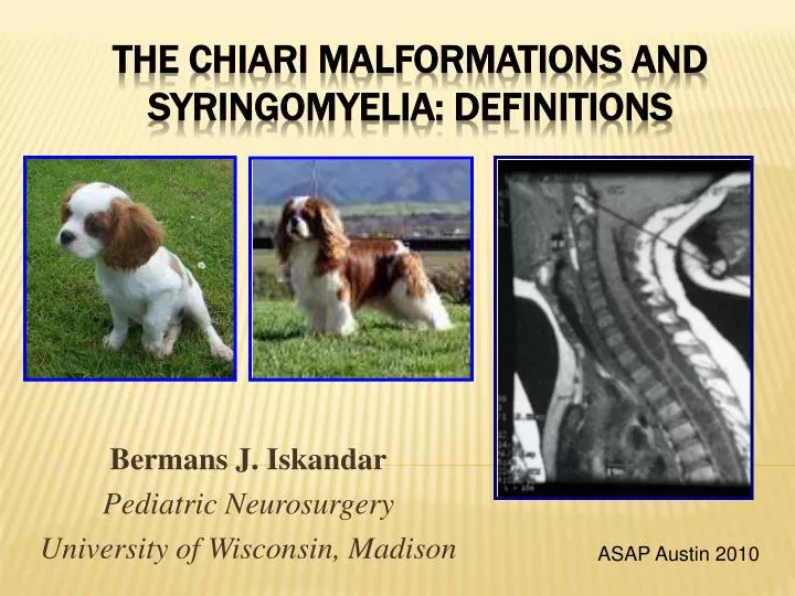
Bermans J. Iskandar Pediatric Neurosurgery University of Wisconsin, Madison. The Chiari Malformations and syringomyelia: Definitions. ASAP Austin 2010. Standard. Chiari Type I Tonsillar descent >5mm below the plane of the foramen magnum.

E N D
Bermans J. Iskandar Pediatric Neurosurgery University of Wisconsin, Madison The Chiari Malformations and syringomyelia: Definitions ASAP Austin 2010
Standard • Chiari Type I • Tonsillar descent >5mm below the plane of the foramen magnum. • No associated brainstem herniation or supratentorial anomalies • Low frequency of hydrocephalus and syringomyelia • Chiari Type II • Caudal descent of the vermis, brainstem, and fourth ventricle. • Associated with myelomeningocele and multiple brain anomalies • High frequency of hydrocephalus and syringohydromyelia
Rare & Poor Prognosis • Chiari Type III • Occipital encephalocele containing • Dysmorphic cerebellar and brainstem tissue • Chiari Type IV • Hypoplasia or aplasia of the cerebellum
New & Controversial • Chiari 1.5 • Descent of tonsils & medulla • Behaves like Chiari I • Chiari Zero • Idiopathic syringomyelia that responds to craniocervical decompression JNS:Peds 2004 JNS 1998
Chiari I Malformation • Diagnosis made on MRI • Treatment: posterior fossa decompression. • If the syrinx does not resolve: • Re-explore the posterior fossa and expand the decompression • Consideration of subtle craniocervical instability • Consideration of benign intracranial hypertension • Consideration of shunting the syrinx directly
Chiari I Case 1: Basic Scenario • 8 year old boy with headaches • Syrinx • 1 cm tonsillar descent
Chiari I Case 2: Pseudotumor Cerebri • 30 year-old with 1.5 cm tonsillar descent and severe symptoms • Posterior fossa decompression fails • LP monitoring reveals elevated ICP • VP shunt • Symptoms resolve
Chiari ICase 4: Acquired Chiari I • 10 year-old who underwent serial lumbar punctures for a mild viral meningitis • Develops lower cranial nerve symptoms • MRI reveals new tonsillar herniation
Chiari I Case 5: Chronic Craniocervical Instability • 12 year-old with Chiari I, syringomyelia, and basilar invagination • Posterior fossa decompression • Symptoms and syrinx don’t resolve until craniocervical fusion a year later
Chiari II Malformation • Likely Etiology • In utero CSF leak through the myelomeningocele opening, causing • caudal traction on brain structures • Clinical Presentation • Infants: usually asymptomatic • Children: signs of lower brainstem compression: stridor, apnea, dysphagia, aspiration
Chiari II Malformation • Chiari II: leading cause of death in spina bifida patients in the recent past • 30% of patients: brainstem symptoms by age 5 (1/3 of these die) • Most dangerous period: 2-3 months of age (sometimes up to 2 years)
Chiari II Malformation • Current understanding • VP shunt malfunction most likely cause of deterioration, rather than the Chiari • Ventricle size may not change • Number of Chiari II decompressions has decreased significantly since more aggressive shunt revisions
Syringomyelia • Fluid-filled cavity within the spinal cord • Other nomenclature • Hydromyelia • Syringohydromyelia • Spinal cord cyst
Signs and Symptoms • Dissociated sensory loss • Central cord syndrome • Brainstem symptoms and signs • Scoliosis • Chronic pain
Diagnostic Studies • Spinal MRIwill show a dilated cavity with the same intensity of CSF. • A complete brain and spinal MRI with and without Gadolinium is needed to determine the primary pathology. • Cine MRImay also help in diagnosing abnormal CSF flow patterns. So far results have been conflicting. • Rarely, myelography may help to sort some of the more difficult cases.
Treatment - Based on Etiology • Asymptomatic patients with small syrinx cavity and no obvious etiology are best managed with watchful waiting and serial imaging • Large syrinx: Treat the cause of the syrinx, not the syrinx itself
Spina Bifida • The syrinx may be the result • Tethered cord from the myelomenigocele repair scar • Chiari II malformation • Ventricular shunt malfunction. • Location of the syrinx within the spinal cord may help to dictate the treatment • Lumbar syrinx ??tethered cord release • Cervical syrinx ?? VP shunt revision • Check the shunt first!
Congenital Tethered Cord (Spina Bifida Occulta) • Diagnosis by MRI • Treatment: Tethered cord release • If syrinx is large, it is often drained at the same surgery
Arachnoiditis • Diagnosis made on MRI • Treatment: Dissection of the arachnoid scar (often difficult or impossible) • Goal: Reestablish normal CSF flow • Difficulties: If the arachnoiditis is so diffuse that it becomes impossible to achieve a good dissection, shunt the syrinx to the pleural or peritoneal cavities
Trauma • Post-traumatic syrinx is difficult to treat successfully • Possible causes • Arachnoiditis and blockage of flow causing expansion of the cord, or • Atrophy long term after cord contusion • Treatment: arachnoidal dissection, or syrinx shunt into the pleura or peritoneum
Spinal Cord Tumor • Diagnosis made on MRI • High protein content • Treatment: • Tumor resection • It is rare to have to shunt the syrinx in these situations.
IdiopathicNo Identifiable Cause • In a large percentage of patients, the syrinx has no identifiable cause • Difficult to treat • If large, syrinx shunting • Rarely, posterior fossa decompression (Chiari zero) • It is so far impossible to predict which patient with idiopathic syringomyelia would respond to posterior fossa decompression
Prognosis and OutcomeSyringomyelia Resolution • Chiari decompression • Excellent outcome • Spina bifida • Excellent outcome when shunt is functional • Arachnoiditis • Focal – fair prognosis • Diffuse – poor, need to shunt the syrinx • Trauma • Poor outcome for syringomyelia and pain • Tumor: • Excellent outcome for syringomyelia • Overall Prognosis depends on tumor grade
Conclusions • Standard basic definitions • Complicating factors: hydrocephalus, pseudotumor cerebri, instability, etc. • Treatment controversies • When to treat • What to do • When to do it • Goals: Recognize basic concepts; recognize controversial areas; be prepared to bring these points up with your physician