Download
1 / 1
10 likes | 133 Views
PJ Mulholland 1 , RL Self 1 , BR Harris 2 , HJ Little 3 , JM Littleton 2 , MA Prendergast 1 1 Department of Psychology, University of Kentucky, Lexington, KY 40506-0044 2 Department of Behavioral Science, University of Kentucky College of Medicine, Lexington, KY 40536-0086

E N D
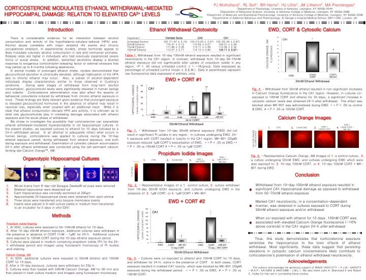
PJ Mulholland1, RL Self1, BR Harris2, HJ Little3, JM Littleton2, MA Prendergast1 1Department of Psychology, University of Kentucky, Lexington, KY 40506-0044 2Department of Behavioral Science, University of Kentucky College of Medicine, Lexington, KY 40536-0086 3Department of Molecular and Biomedical Pharmacology, University of Kentucky College of Medicine, Lexington, KY 40536-0298 4Departments of Addictive Behaviour and Pharmacology, St George's Hospital Medical School, SW17 ORE, London, UK CORTICOSTERONE MODULATES ETHANOL WITHDRAWAL-MEDIATED HIPPOCAMPAL DAMAGE: RELATION TO ELEVATED CA2+ LEVELS CA1 CA3 DG Introduction Ethanol Withdrawal Cytotoxicity EWD, CORT & Cytosolic Calcium There is considerable evidence for an interaction between alcohol consumption and activity of the hypothalamic-pituitary-adrenal (HPA) axis. Alcohol abuse correlates with major stressful life events and chronic occupational stressors. In experimental models, stress hormones appear to likely modulate voluntary alcohol consumption in rats and nonhuman primates. Relapse rates are higher in individuals who had previously experienced severe forms of social stress. In addition, detoxified alcoholics display a blunted response to exogenous corticotrophin releasing factor or external stressors that may persist up to 6 months following abstinence. In animal models of prolonged ethanol intake, studies demonstrated that glucocorticoid secretion is chronically elevated, although habituation of the HPA axis to chronic ethanol may occur. Also, a subset of alcohol-dependent individuals display characteristics similar to those observed in Cushing’s syndrome. During early stages of withdrawal from long-term alcohol consumption, glucocorticoid levels were significantly elevated in human beings and rodents. Corticosterone administration may also affect the severity of behavioral convulsions induced by withdrawal from chronic ethanol exposure in mice. These findings are likely relevant given evidence that prolonged exposure to elevated glucocorticoid hormones in the absence of ethanol may result in neuronal loss, especially when coupled with an additional insult. While it is clear that ethanol consumption disrupts HPA axis activity, it is unknown as to what role glucocorticoids play in mediating damage associated with ethanol exposure and the acute phase of withdrawal. We chose to investigate the possibility that corticosterone can exacerbate ethanol withdrawal-mediated hyperexcitability in rat hippocampal cultures. In the present studies, we exposed cultures to ethanol for 10 days followed by a 24-h withdrawal period. In an attempt to adequately reflect what occurs in human beings, corticosterone was applied to cultures during the 10-day ethanol exposure period, during withdrawal from ethanol exposure, and both during exposure and withdrawal. Examination of cytosolic calcium accumulation 24-h after ethanol withdrawal was conducted using the cell permeant calcium binding dye Calcium OrangeTM, AM. Dentate Gyrus CA3 CA1 Treatment Untreated Control 100 (11.87 ± 2.74) 100 (13.03 ± 3.07) 100 (20.59 ± 2.87) 18 * CA1 50mM Ethanol 110.41 ± 3.13 112.63 ± 5.07 109.51 ± 5.48 16 75mM Ethanol 111.89 ± 3.39 113.11 ± 5.99 110.08 ± 5.56 100mM Ethanol 112.57 ± 3.77 114.15 ± 4.80 125.11 ± 3.97* 14 12 # Table 1.Withdrawal from 10-day 100mM ethanol exposure resulted in significant neurotoxicity in the CA1 region. In contrast, withdrawal from 10-day 50-75mM ethanol exposure did not significantly alter uptake of propidium iodide in any region. *P < .05 versus untreated control. n = ~18/group. Data expressed as percentage of untreated control (mean ± S.E.M.). Data in parentheses represent raw fluorescence data expressed in arbitrary units. CALCIUM ORANGE, AM FLUORESCENCE 10 8 6 4 2 0 EWD + CORT #1 100nM CORT+ EWD CORT MK-801 50mM Ethanol Withdrawal 50 ** CA1 Fig. 4. – Withdrawal from 50mM ethanol resulted in non-significant increases in Calcium Orange fluorescence in the CA1 region. However, in cultures co-exposed to 100nM CORT and ethanol for 10-days, a significant increase in cytosolic calcium levels was observed 24-h after withdrawal. This effect was blocked when MK-801 was administered during EWD. * = P < .05 vs control & EWD, # = P < .05 vs 100nM CORT. 40 * HIPPOCAMPAL DAMAGE (uptake of propidium iodide) 30 20 # 10 Calcium Orange Images 0 +MK-801 EWD 1nM 10nM 100nM 1mM 24 hr CORT 50mM Ethanol Withdrawal EWD + 100nM CORT + MK-801 EWD + 100nM CORT Fig. 1. – Withdrawal from 10-day 50mM ethanol exposure (EWD) did not result in significant PI uptake in any region. In cultures undergoing EWD, 24-h exposure with CORT resulted in toxicity in the CA1 region. MK-801 (20mM) exposure reduced 1mM CORT’s exacerbation of EWD. * = P < .05 vs EWD,** = P < .05 vs 100nM CORT # = P < .05 vs 1mM CORT. 1. 2. 3. 4. CTRL 50mM EWD Elevated CALCIUM ORANGE FLUORESCENCE Propidium Iodide Images Fig. 5. – Representative Calcium Orange, AM images of 1. a control culture, 2. a culture undergoing 50mM EWD, and cultures undergoing EWD which were also exposed to: 3. 10-day 100nM CORT, or 4. 10-day 100nM CORT + MK-801 during EWD. Organotypic Hippocampal Cultures EWD + 1mM CORT + MK-801 EWD + 1mM CORT 1. 2. 3. 4. CTRL 50mM EWD Damaged PI UPTAKE 2. 3. 4. 5. 1. 6. Conclusion Withdrawal from 10-day 100mM ethanol exposure resulted in significant CA1 hippocampal damage as opposed to withdrawal from 50-75mM ethanol exposure Marked CA1 neurotoxicity, in a concentration-dependent manner, was observed in cultures exposed to CORT during 50mM ethanol exposure and/or withdrawal When co-exposed with ethanol for 10 days, 100nM CORT was associated with elevated Calcium Orange fluorescence (~16% above controls) in the CA1 region 24-h after withdrawal Fig. 2. – Representative images of a 1. control culture, 2. culture withdrawn from 10-day 50mM EtOH exposure, and cultures undergoing EWD in the presence of: 3. 1mM CORT, or 4. 1mM CORT + MK-801. 1.Whole brains from 8-day-old Sprague Dawley® rat pups were removed 2.Bilateral hippocampi were dissected out 3.Each hippocampus was coronally sectioned at 200mm 4.Approximately 24 hippocampal slices were obtained from each animal 5.Three slices were transferred onto biopore membrane inserts 6.Inserts were placed in 6-well culture plates in medium then transferred to an incubator for 5 days in vitro (DIV) EWD + CORT #2 180 CA1 * Methods 160 140 * 120 Propidium Iodide Staining 1. At 5DIV, cultures were exposed to 50-100mM ethanol for 10 days. 2. After 10-day 50mM ethanol exposure, additional cultures were withdrawn in the presence or absence of CORT (1nM – 1mM) for 24-h. Additional cultures were exposed to 100nM CORT during the 10-day ethanol exposure period. 3.Cultures were placed in medium containing propidium iodide (PI) for the 24-h withdrawal period and imaged using fluorescent microscopy of PI nucleic acid staining. HIPPOCAMPAL DAMAGE (uptake of propidium iodide) 100 80 60 40 Thus, this study demonstrates that corticosterone exposure sensitizes the hippocampus to the toxic effects of ethanol withdrawal. Most significantly, these data suggest that persisting elevations in cytosolic calcium concentrations likely contribute to corticosterone’s potentiation of ethanol withdrawal neurotoxicity. # # 20 0 +MK-801 +MK-801 EWD CORT EWD CORT 10-day CORT 10-day + WD CORT 50mM Ethanol Withdrawal Calcium Orange, AM 1. At 5DIV, additional cultures were exposed to 50mM ethanol and 100nM CORT for 10 days. 2. After a 10-day exposure, cultures were withdrawn for 23½-h. 3. Cultures were then loaded with 500nM Calcium Orange, AM for 30-min and then placed in fresh culture medium and imaged using fluorescent microscopy Fig. 3. – Cultures were co-exposed to ethanol and 100nM CORT for 10 days, and withdrawn for 24 hr, some in the presence of CORT. In both cases, CORT exposure resulted in marked CA1 toxicity, which was blocked by MK-801 (20mM) exposure during the withdrawal period. * = P < .05 vs EWD, # = P < .05 vs 100nM CORT. Acknowledgements The authors acknowledge the generous support of NIAAA (AA014771 – P.J.M.; AA00274 – M.A.P.; AA12600 & AA013388 – J.M.L.).We also thank John A. Blanchard II and Robert C. Holley for their aid in completing these studies.