Download
1 / 52
550 likes | 722 Views
Ling 411 – 22. The Functional Neuroanatomy of Language (Part II). Applying the findings about columnar organization. Christine Cooper:

E N D
Ling 411 – 22 The Functional Neuroanatomy of Language (Part II)
Applying the findings about columnar organization Christine Cooper: I have recently been thinking a lot about the broader implications, and even potentially applications, of all of the knowledge on columnar organization. It seems to me like this information could be invaluable to childcare and education.
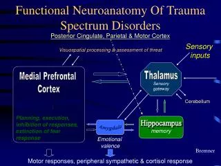
http://www.pnas.org/content/97/22/11850.full.pdf+html Patricia Kuhl on learning perceptual distinctions:An example A Fig. 6. (A) Physical distance between /ra-la/ syllables in a grid created by varying formants 2 and 3 in equal steps. (B) Perceptual distance between syllables for American listeners. B
Ling 411 – 22 The Functional Neuroanatomy of Language (Part II)
Some earlier findings w.r.t. RH speech perception • Vowel qualities • Intonation • Tones in tone languages
Imaging studies • When listening to spoken discourse, cerebral blood flow increases in • Wernicke’s area • Broca’s area • RH homologues of Wernicke’s and Broca’s areas • More cerebral blood flow in RH when subjects read sentences containing metaphors than literal sentences
Experiments (described by Beeman) • Words presented to rvf-LH or lvf-RH • RH more active than LH • Synonyms • Co-members of a category: table, bed • Polysemy: FOOT1 – FOOT2 • Metaphorically related connotations • Sustains multiple interpretations • LH about same as RH • Other associations: baby-cradle • LH more active than RH • Choose verb associated with noun
Patients with brain-damage • Some patients with LH damage • Can’t name fruits but can say that they are fruits • Patients with RH damage • Impaired comprehension of metaphorical statements • More difficulty producing words from a particular semantic category than producing words beginning with a particular letter (258)
Experiments on speech perception • Dichotic listening – normal subjects • Right ear (i.e. LH) advantage for distinctions of • Voicing • Place of articulation • Left hear (RH) advantage for • Emotional tone of short sentences • Sentences presented in which only intonation could be heard • RH advantage for identifying sentence type – declarative, question , or command
Experiments on speech perception • Split brain patients • They hear a consonant • Then written representations are presented • ‘Point to the one you heard’ • rvf-LH exhibited strong advantage
Patients with right-brain damage • Posterior RH lesions result in deficits in interpreting emotional tone • Anterior RH lesions abolish the ability to control the production of speech intonation
Split-brain studies • Isolated RH has ability to read single words • But not as fast nor as accurate as LH • Ability declines with increasing word length • Lexical context does not assist letter identification • In Japanese subjects • RH is better at reading kanji than kana • LH is better at reading kana
RH involvement in speech perceptionEvidence from an earlier paper by Hickok • Evidence from tests of isolated RH • Split-brain studies • Wada test • Sodium amytol, sodium barbitol • Discrimination of speech sounds • Comprehension of syntactically simple speech (Hickok 2000: 92)
Caution – Split-Brain Studies • These patients are generally epileptics • Usually the onset of seizures is several to many years before the surgery • Often the onset of seizures was during childhood • Therefore the brain has had time to adapt – perhaps reorganize some linguistic functions
RH involvement in speech perceptionIntra-operative recording • Evidence from intraoperative recording • Sites found in STG of both hemispheres for • Phoneme clusters • Distinguishing speech from backwards speech • Distinguishing mono- from polysyllabic words (Hickok 2000: 92-3)
RH involvement in speech perceptionImaging • Evidence from imaging • PET • fMRI • MEG • Subjects passively listen to speech • Both hemispheres show activity • More activity in LH • Some evidence for differential contributions of the two hemispheres (Hickok & Poeppel, another publication) (Hickok 2000: 93)
5. Phonological processing systems in speech recognition are bilateral but asymmetric • Hickok: “The hypothesis that phoneme-level processes in speech recognition are bilaterally organized does not imply that the two hemispheres are computationally identical. • “In fact there is strong evidence for hemispheric differences in the processing of acoustic/speech information [2,17,68,90,193].” • Basis of the differences? – Two views • Temporal (LH) vs. spectral (RH) resolution • Difference in sampling rate • LH: 25-50 Hz • RH: 4-8 Hz
Another opportunity for either-or thinking:Explaining differential functions of LH and RH (127) • One possibility: • Temporal vs. spectral resolution • Another possibility (Zatorre): • Different sampling rates • LH – 25-50 Hz • RH – 4-8 Hz • It’s not either-or!
Fig. 5. Serial vs. parallel models of speech recognition “The processing levels may be distributed across the two hemispheres in some fashion and may correspond to different temporal windows of integration”
Possible bases for RH/LH difference • Higher ratio of white to gray matter in RH • Therefore, higher degree of connectivity in RH • Difference in dendritic branching • Different density of interneurons • Evoked potentials (EEG) are more diffuse over the RH than over LH Beeman 257
Grammatical and semantic/conceptual information • There’s a lot we don’t know • Hickok: “The neural organization of conceptual-semantic systems is a matter of debate” (127) • What we do know: • Lexico-grammatical and semantic-conceptual – 2 levels • Semantic-conceptual is very widely distributed • Different areas for different categories (cardinal noses) • Areas commonly implicated: • Posterior MTG and ITG • Also, other temporal areas • AG
Hickok’s Fig. 1 Hickok: MTG/ITG (LH and RH) are “important in mapping sound onto meaning” ATL (LH) implicated by some in “lexical-semantic and sentence-level processing”
7. Posterior language cortex in the left hemisphere is involved in phonological aspects of speech production • Hickok gets it right: Wernicke’s area is heavily involved in speech production • Provides additional evidence (q.v. – 128, 129) • “Given these behavioral observations, it is no surprise that posterior sensory-related cortex in the left hemisphere have been found to play an important role in speech production.” (129)
Wernicke’s area in speech production “What I would like to suggest is that Wernicke was essentially correct in hypothesizing … that auditory cortex participates in speech production …” (Hickok 2000: 89)
Hickok quotes Wernicke: Observations of daily speech usage and the process of speech development indicates the presence of an unconscious, repeated activation and simultaneous mental reverberation of the acoustic image which exercises a continuous monitoring of the motor images. Wernicke 1874
Evidence for left pSTP* involvement in speech production • Erratic speech of Wernicke’s aphasics • Conduction aphasia from damage to left pSTP* • Intraoperative stimulation of left pSTP* • “distortion and repetition of words and syllables” (Penfield & Roberts 1959) • N.B.: As in Wernicke’s aphasia • MSI study shows activity in left pSTG just before speech production (picture naming) (Levelt et al. 1998) • fMRI study: similar results – no RH activity shown (Hickok et al. 1999) (Hickok 2000: 93-4) *pSTP: posterior Supra-Temporal Plane
Spt: active in both perception and production (Fig. 8A) “A number of fMRI studies have demonstrated the existence of an area in the left posterior Sylvian region (area Spt, Fig. 8A) that responds both during the perception and production of speech (Fig. 8B), even when speech is produced covertly (subvocally)…” (130) “Conduction aphasics…typically have damage involving Spt” (132)
Area “Spt” (proposed by Hickok & Poeppel) “We occasionally get questions regarding how to define area Spt -- the key dorsal-stream region we believe performs sensory-motor transformations for speech. The acronym stands for Sylvian parietal temporal to reflect the fact that it is located within the Sylvian fissure at the parietal-temporal boundary. The region involves a portion of the planum temporal/parietal operculum (very hard to distinguish the two), and is a subportion of area tpt.” http://www.talkingbrains.org/2007/05/where-is-area-spt.html Seems to overlap with the TPO junction area and/or SMG
Area “Spt” in an fMRI experiment http://www.talkingbrains.org/search/label/commentary
Area Spt not just for speech Also involved in music perception and production (humming)
Subdivisions of Planum temporale (Fig. 6) “Note that there are four different fields within the planumtemporale suggesting functional differentiation, and that these fields extend beyond the planumtemporale”
Problem with Hickok’s proposal • Hickok proposes that • phonological recognition is bilateral • but conduction aphasia results from Spt in LH • Problem: When patients are given words to repeat • Conduction aphasics keep trying • Wernicke aphasics don’t • Indicates that Wernicke aphasics don’t perceive their own speech • Another problem: Wernicke aphasia usually results from LH damage only
Repetition in Wernicke’s aphasia Model for Repetition Patient’s Response blackboard shoelace He park … he came with the car. He came with his car. It went two cars … between the cars • black • shoe • He parks the car • It goes between two others
Picture naming in conduction aphasia • Picture of.. • Patient’s Response • tris.. chi.. trissle.. sissle.. twiss.. ciss. • trep.. tretzle.. trethle.. tredfl… ki • whistle • pretzel
Lamb’s email query to Hickok (April 1, 2010) Hi Greg - In my neurolinguistics class we have just been considering your 2000 paper from the Grodzinsky et al. volume, with its new perspective on, among other things, these two types of aphasia. Very intriguing, but I have a question: How do you explain this: When you give a conduction aphasic words to repeat, he/she commonly produces a phonemic paraphasia and then keeps trying, since he/she recognizes the error; but a Wernicke's aphasic usually stops after one incorrect repetition, evidently unaware of the error. Acc. to your proposal, both types of aphasic have LH phonological recognition wiped out, and both have intact RH pSTG.
Hickok’s response (April 1, 2010) Hi Syd, Good to hear from you. That is an interesting question. I think there are two possibilities. One is that the conduction aphasics don't have as much damage to the left hemisphere phonological systems we (I) might have thought. I.e., the damage is more often involving the posterior Sylvian region (Spt). The intact left and right hemi phonological systems allow the patient to clearly recognize their errors and self correct. Wernicke's on the other hand typically have extensive damage to the left hemi phonological systems which, because of their role in production, may have a larger role in self monitoring. Another possibility, perhaps in conjunction with the first, is that Wernicke's have damage to semantic (access) systems as well. it may be much harder to notice a phonological error if you can't tell whether it is a word or not.
The Perception-Production InterfaceAlternative views • We have phonological recognition in Wernicke’s area • And in RH homolog of Wernicke’s area • And phonological production in Broca’s area • And in RH homolog of Broca’s area • Clearly, they have to be connected • The traditional view • Direct connection: the arcuate fasciculus • Proposed by Wernicke, supported by Geschwind • Alternative view • Supramarginal gyrus (SMG) as intermediary • Proposed by Hickok • with support from Damasio and Goldstein
The Intermediate System Hypothesis(Two versions) Auditory-Motor Interface Speech Production Speech Recognition SMG (proposed by some) Hickok’s alternative: Spt
Arguments for the Direct Connection Hypothesis • No additional intervening structure needed • We have anatomical evidence for the arcuate fasciculus • And for its connections from Wernicke’s area to Broca’s area
Arguments for the intermediate system hypothesis • Damasio cites SMG damage as a major cause of conduction aphasia • Consistent with earlier findings of Goldstein • “Central aphasia” (Goldstein 1948) • Connectivity studies in non-human primates fail to find direct connection between auditory cortex and ventral posterior frontal lobe • But support the claim that the lower parietal lobe provides an interface between these areas (Hickok 2000: 99) • SMG is a likely site of higher-level proprioceptive processing of speech
Arguments for the intermediate system hypothesis • Damasio cites SMG damage as a major cause of conduction aphasia • Consistent with earlier findings of Goldstein • “Central aphasia” (Goldstein 1948) • Anatomical studies in macaque monkeys fail to find direct connection (between corresponding areas) (Hickok 2000: 99) • SMG is a likely site of higher-level proprioceptive processing of speech • (next slide)
Motor and Somatosensory Areas for speech Central Sulcus Post-Central Sulcus 1-Phonological production 2-Articulation 3-Articulatory monitoring 4-Phonological monitoring Leg Trunk Arm 4 Hand Fingers 3 1 2 Mouth
Presumed interconnections of speech areas Central Sulcus Post-Central Sulcus 1 – Phonological production 2 – Articulation 3 – Articulatory monitoring 4 – Phonological monitoring 5 – Primary auditory 6 – Phonological recognition 4 3 1 2 6 5
And there’s more than meets the eye • The phonological recognition area includes the temporal plane • The phonological monitoring area includes the parietal operculum • Both very large areas
REVIEW The Sylvian Fissure
REVIEW Evidence for left pSTP involvement in speech production • Erratic speech of Wernicke’s aphasics • Conduction aphasia from damage to left pSTP • Intraoperative stimulation of left pSTP • “distortion and repetition of words and syllables” (Penfield & Roberts 1959) • N.B.: As in Wernicke’s aphasia • MSI study shows activity in left pSTG just before speech production (picture naming) (Levelt et al. 1998) • fMRI study: similar results – no RH activity shown (Hickok et al. 1999) (Hickok 2000: 93-4)
Diffusion Tensor Imaging (DTI) • New and very informative technique • Uses MRI • Allows observation of molecular diffusion in living tissues • Makes use of • Brownian movement • Magnetic properties of hydrogen nuclei • Two of them in every water molecule (H2O) • Water moves along lines of least resistance • i.e., along white matter axons • aided by myelin
Arcuate fasciculus in primates Asif Ghazanfar, Nature Neuroscience 11:4.382-384, April 2008
Uniformity of cortical strucureacross mammals? • The hypothesis of uniformity • Very important for perceptual neuroscience • Allows data from experiments on cats and monkeys to be applied to human cortical structure and function • Including higher levels – language • But: this hypothesis applies to grey matter • Not white matter • Cortico-cortical connections • DTI shows that white matter connections differ across mammals
Arcuate fasciculus in different primates Asif Ghazanfar, Nature Neuroscience 11:4.382-384, April 2008