Download
1 / 24
250 likes | 396 Views
DRUG PERMEATION THROUGH SKIN. Mario Grassi Department of Chemical Engineering (DICAMP) UINVERSITY OF TRIESTE. non-living layer of keratin-filled cells surrounded by a lipid-rich extracellular matrix. viable tissue devoid of blood vessels. contains capillary loops. 1 - SKIN STRUCTURE.

E N D
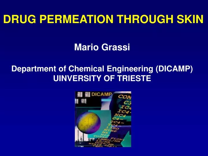
DRUG PERMEATION THROUGH SKIN Mario Grassi Department of Chemical Engineering (DICAMP) UINVERSITY OF TRIESTE
non-living layer of keratin-filled cells surrounded by a lipid-rich extracellular matrix viable tissue devoid of blood vessels. contains capillary loops 1 - SKIN STRUCTURE Adapted from M. Prausnitz et al. NATURE REVIEWS, DRUG DISCOVERY, 3, (2004),115
Hair 1 2 3 Stratum Corneum Sweat pore Epidermis Sweat duct Sebaceous gland Sweat gland Hair follicle Dermis 2 – PERMEATION ROUTES Adapted from B. W. Barry, Adv. Drug. Del. Review, 54, (2002), S31-40
4 – MODELLING1 Cd = Cd0 M = M0 Drug DRUG DISSOLUTION Css = 0 Cde = 0 DRUG DIFFUSION Css = 0 DRUG CONCENTRATION INCREASE Cr = 0
FICK LAW S. Corneum Dermis + Epidermis stagnant layer
SOLID SURFACE VARIATION: MONODISPERSED PARTICLES SYSTEM SOLID DRUG Mass balance Particles initial surface area
SAMPLING TECNIQUE receiver drug concentration just before sampling. receiver drug concentration just after sampling.
CASE STUDY: ACYCLOVIR PERMEATION THROUGH RAT SKIN ACYCLOVIR - Recordati , Milano -ANTIVIRAL, WHITE CRYSTALLINE POWDER (R = 5.7 mm) -SURFACE AREA = 3370 cm2/g (mercury porosimeter) -U.V. PEAK ABSORBANCE 251 nm - SOLUBILITY in PBS (pH = 7.4; 37°C) Cs = 2.62 mg/cm3 - ACYCLOVIR DIFF. COEFF. (PBS, 37°C) Dss = 7.8*10-6 cm2/s (IDR) - ACYCLOVIR DISS. CONST. (PBS, 37°C) KD = 5*10-5 cm/s • RAT SKIN: • Male hairless rats (Rnu eutimic, Charles River, MI, Italy) 5–7 weeks old • Full-thickness skin removed from abdomen by incision of the outermost layer with a surgical bisturi. Stratum corneum was separated from the dermis–epidermis by placing the full-thickness skin (dermis-side down) on a filter paper saturated with a 1% trypsin) solution at the temperature of 37 ◦C for 4 h.
STAGNANT LAYER THICKNESS: hss Sh = f Rem Scn Sh = d/hss Sc = h/(r D) Re = wrd2/h r = fluid density D = drug diff. coeff. d = stirrer diameter h = fluid viscosity. Dissolution w = 388 rpm w = 505 rpm w = 605 rpm hss1 ==> Sh1 hss2 ==> Sh2 hss3 ==> Sh3 T = 25°C w = 388 rpm w = 505 rpm w = 605 rpm hss4 ==> Sh4 hss5 ==> Sh5 hss5 ==> Sh6 T = 37°C
f = 3.2*10-6 m = 1.2 n = 0.95 Sc = 1147 Re = 5737 hss = 0.011 cm
PARTITION COEFFICIENTS: Kp1,2,3,4 The full skin/Acyclovir solution and the one-layer skin/Acyclovir solution partition coefficients Kskin and Kde are determined by immersion of both fragments in the Acyclovir solution (PBS pH 7.4) for 4 h at 37°C Kskin =0.547 Kde =0.95 Kp1 =0.5 (literature) Kp3 = Kp2*Kp1 Kp2 = Kskin*(1+G)-Kp1/(G*Kp1) G = hsd/hsc= 11.7 Kp1 = 0.5 Kp2 = 1.1 Kp4 = 1 Kp3 = 0.55
5 – RESULTS FIVE WEEKS OLD RATS: ONE LAYER (DERMIS-EPIDERMIS) Data (symbols) not corrected for dilution D = 2*10-6 cm2/s Lines: model best fitting D = 2*10-6 cm2/s D = 1*10-6 cm2/s DAV = (1.7 ± 0.6)*10-6 cm2/s
SEVEN WEEKS OLD RATS: ONE LAYER (DERMIS-EPIDERMIS) Data (symbols) not corrected for dilution D = 9.5*10-7 cm2/s Lines: model best fitting D = 6.5*10-7 cm2/s DAV = (8 ± 2)*10-7 cm2/s
FIVE WEEKS OLD RATS: FULL SKIN D = 2.5*10-9 cm2/s Data (symbols) not corrected for dilution Lines: model best fitting D = 1*10-9 cm2/s D = *10-10 cm2/s DAV = (1.33 ± 1)*10-9 cm2/s
SEVEN WEEKS OLD RAT: FULL SKIN D = 6.5*10-10 cm2/s Data (symbols) not corrected for dilution Lines: model best fitting
COMMENTS Regardless animal age, stratum corneum represents the main barrier to drug permeation (Dde 1000 Dsc) 1 2 3 4 Skin permeability seems to decrease with age A considerable inter-animal variability is observed Model simulations reveal that pseudo steady state conditions are met after 2 hours for young animals (one layer skin), while they are met after 6 hours in the remaining cases
6 – COMPARISON THIS APPROACH / COMMON APPROACH 2 = corrected drug concentration after “n” samplings = experimental drug concentration after “n” samplings = experimental drug concentration after “i” samplings = sampling volume = receiver volume DATA CORRECTION FOR DILUTION
ASYMPTOTIC FICK EQUATION SOLUTION 1) Pseudo steady state conditions 2) Cd = C0; Cr 0 (sink conditions) 3) Stagnant layer is neglected Dm = drug diff coeff through membrane h = membrane thickness S = membrane area Kp = partition coefficient
ASSUMING “m” AND “q” AS INDEPENDENT FITTING PARMETERS: IS FITTED ON EXPERIMENTAL DATA TO GET Dm
FROM DIFFUSION COEFFICIENT TO RESISTANCE TRADITIONAL APPROACH THIS APPROACH
Experiment number identifier N Animal age (weeks) F-value Ro (s/cm) Rm (s/cm) Dermis + epidermis (one layer skin) 1 5 732 (5.9 0.014)*104 (6.1 0.016)*104 2 5 261 (4.9 0.001)*104 (5.5 0.012)*104 3 5 136 (12.6 0.06)*104 (12.1 0.059)*104 4 7 864 (21.1 1.62)*104 (19.2 1.35)*104 5 7 5906 (15.6 0.814)*104 (15.0 0.765)*104 Full skin 6 5 595 (4.83 0.75)*106 (2.49 0.175)*106 7 5 45 (26.4 24.7)*106 (10.7 2.96)*106 8 5 99965 (13.7 6.7)*106 (7.68 0.014)*106 9 7 57 (12.7 5.2)*106 (5.76 0.13)*106 This approach Traditional approach
7 – REFERENCES 1) N. Coceani, I. Colombo, M. Grassi, Int. J. Pharm. 254 (2003) 197 –210. 2) Chien, Y.W. (Ed.), 1987. Transdermal Controlled Systemic Medications.Marcell Dekker, Inc., New York, Basel (Chapter 2).