Download
1 / 21
290 likes | 568 Views
REGULATION OF CELL CYCLE BY PROTEIN KINASES. BIOL 306 -BIOCHEMISTRY II H.ESRA AKGÜL 01040604. References:. LEHNINGER PRINCIPLES OF BIOCHEMISTRY www.biop.ox.ac.uk www.biochemj.org. SUBJECTS:. AN INTRODUCTION CELL CYCLE IMPORTANCE OF PROTEIN KINASES

E N D
REGULATION OF CELL CYCLE BY PROTEIN KINASES BIOL 306 -BIOCHEMISTRY II H.ESRA AKGÜL 01040604
References: • LEHNINGER PRINCIPLES OF BIOCHEMISTRY • www.biop.ox.ac.uk • www.biochemj.org
SUBJECTS: • AN INTRODUCTION • CELL CYCLE • IMPORTANCE OF PROTEIN KINASES • LEVELS OF CYCLIN DEPENDENT PROTEIN KINASES OSCILLATES • CRITICAL PROTEINS IN CDKs REGULATE
Cell division requires a ordered sequence of biochemical events that assures every daughter cell a full complement of molecules required for life. Control of cell division in eukaryotic cells have revealed regulatory mechanisms. Protein kinases and protein phosphorylation are central to the timing mechanism that determines entry into cell division and ensures orderly passage through
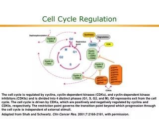
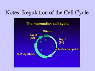
Phases: • S PHASE:DNA is replicated to produce copies for both daughter cells. • G2 PHASE:New proteins are synthesize and the cell double in size • M PHASE: (MITOTIC)maternal nuclear envelope breaks down,paierde chromosome are pulled to opposire of cell and cytokinesis ,producing two daughter cell • G1 PHASE:The waiting period of again dividing. • G0 PHASE: Ceases phase,entering the quiscent.If cell starts to divide,it reenters to G1 .
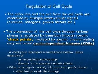
IMPORTANCE OF PROTEIN KINASES • The timing of the cell cycle is controlled by protein kinases with activities that change in response to cellular signals. • By phosphorylating of proteins,protein kinases the metabolic activities to produce cell division.
WHAT IS CDKs? • The kinases are heterodimers with a regulatory subunit, cyclin, and a catalytic subunit, cyclin-dependent protein kinase (CDK). In the absenceof cyclin, the catalytic subunit is virtually inactive.When cyclin binds, the catalytic site opens up, aresidue essential to catalysis becomes accessible
CDK activities show striking oscillations These oscillations are the result of four mechanisms for regulating CDK activity: 1-phosphorylation or dephosphorylation of the CDK, 2- controlled degradation of the cyclin subunit, 3-periodic synthesis of CDKs and cyclins, 4-the action of specific CDKinhibiting proteins.
Regulation of CDks by phosphorylation The activity of a CDK is strikingly affected by two critical residues in the protein Phosphorylation of Tyr15 near the amino terminus renders CDK2 inactive; the P –Tyr residue is in the ATP-binding site of the kinase, and the negatively charged phosphate group blocks the entry of ATP. A specific phosphatase dephosphorylates this P –Tyr residue, permitting the binding of ATP. Phosphorylation of Thr160 in the “T loop” of CDK, forces the T loop out of the substrate binding cleft, permitting substrate binding and catalytic activity.
Controlled degradation of cyclin: • Progress through mitosis requires first the activation then the destruction of cyclins A and B, which activate the catalytic subunit of the M-phase CDK. These cyclins contain near their amino terminus the sequence Arg–Thr–Ala–Leu–Gly–Asp–Ile–Gly–Asn, the “destruction box,” which targets them for degradation.The DBRP(destruction box recognizing protein.) recognize this protein.
Ubiquitin:Cyclin and activated ubiquitin are covalently joined by the enzyme ubiquitin ligase Several more ubiquitin molecules are then appended, providing the signal for a proteolytic enzyme complex, or proteasome, to degrade cyclin.
The feedback mechanism • Increased CDK activity activates cyclin proteolysis. Newly synthesized cyclin associates with and activates CDK, which phosphorylates and activates DBRP. Active DBRP then causes proteolysis of cyclin. Lowered [cyclin] causes a decline in CDK activity, and the activity of DBRP also drops through slow, constant dephosphorylation and inactivation by a DBRP phosphatase.The cyclin level is ultimately restored by synthesisof new cyclin molecules.
Regulated Synthesis of CDKs and Cyclins • CyclinD, cyclin E, CDK2, and CDK4 are synthesized only when a specific transcription factor, E2F, is present in the nucleus to activate transcription of their genes. Synthesis of E2F is in turn regulated by extracellular signals such as growth factors and cytokines
Inhibition of CDKs • Specific protein inhibitors bind to and inactivate specific CDKs. One such protein is p21.
How does the activity of CDK control the cell cycle? BehindCDK regulation by inspecting the effect of CDKs on the structures of laminin and myosin and on the activity of retinoblastoma protein. pRb; when DNA damage is detected, this protein participates in a mechanism that arrests cell division in G1
Breakdown of the nuclear envelope before segregation of the sisterchromatids in mitosis is partly due to the phosphorylationof laminin by a CDK, which causes laminin filaments to depolymerize. • After the division, CDK phosphorylates a small regulatory subunit of myosin, causing dissociation of myosin from actin filaments and inactivating the contractile machinery
When pRb, is phosphorylated,itcannot bind and inactivate EF2, a transcription factor that promotes synthesis of enzymes essential to DNA synthesis. If the regulatory protein p53 is activated by ATM and ATR, protein kinases that detect damaged DNA, it stimulates the synthesis of p21, which can bind to and inhibit cyclin E–CDK2 and thus prevent phosphorylation of pRb. Unphosphorylated pRb binds and inactivates E2F, blocking passage from G1 to S until the DNA has been repaired.