Download
1 / 36
380 likes | 709 Views
Pathohysiology of ascites. Waleed Al- hamoudi. Ascites. Ascites is of Greek derivation ("askos") and refers to a bag or sack and describes pathologic fluid accumulation within the peritoneal cavity. Most patients (85%) with ascites have cirrhosis.

E N D
Pathohysiology of ascites Waleed Al-hamoudi
Ascites • Ascites is of Greek derivation ("askos") and refers to a bag or sack and describes pathologic fluid accumulation within the peritoneal cavity. • Most patients (85%) with ascites have cirrhosis. • The most common causes of cirrhosis at the present time are chronic viral hepatitis and alcoholic liver disease.
Peritoneal cavity • It is a potential space between the parietal peritoneum and visceral peritoneum, the two membranes separate the organs in the abdominal cavity from the abdominal wall. • Derived from the coelomic cavity of the embryo. • Largest serosal sac in the body and secretes approximately 50 ml of fluid per day.
Peritoneal fluid • It is a normal, lubricating fluid found in the peritoneal cavity. • The fluid is mostly water with electrolytes, antibodies, white blood cells, albumin, glucose and other biochemicals. • Reduce the friction between the abdominal organs as they move around during digestion.
Ascites • Cirrhosis • Infection (TB) • Malignancy • CHF • Nephrotic syndrome • Pancreatic or biliary ascites
Pathogenesis • 1-Increased hydrostatic pressure • 2-Decreased colloid osmotic pressure • 3-Increase in the permeability of peritoneal capillaries • 4-Leakage of fluid into the peritoneal cavity • 5-Miscellinious
Pathogenesis • Cirrhotic Ascites : The most recent theory of ascites formation, the "peripheral arterial vasodilation hypothesis," . This happens as a consequence of portal hypertension.
Introduction • Hepatic blood flow is normally about 1500 mL/minute. • Normal, uncorrected pressure in the portal vein ranges from 5 to 10 mm Hg. Gradient of 2-6. • Portal HPN present when gradient > 12 mmHg. • Approximately 2/3 of the hepatic blood supply is provided by portal venous blood. • The high-pressure, well-oxygenated hepatic arterial blood mixes completely with the low-pressure, low-oxygen-containing, nutrient-rich portal venous blood within the hepatic sinusoids.
The sinusoids are normally protected from upstream portal perfusion pressure and fluctuations. Because they are lined by an endothelium contains a multitude of large (50 to 200 nm), highly permeable fenestrae. • Another feature is hepatic arterial buffer response and is an adenosine-mediated vascular reflex.
After perfusing the sinusoids, blood flows into the hepatic venules, hepatic veins, and inferior vena cava. • Normal hepatic sinusoidal microcirculation has low perfusion pressure which is attributed to the unusually high precapillary to postcapillary resistance in the liver.
Pathophysiology and Causes • The pathogenesis of portal hypertension involves the relationship between portal venous blood flow and the resistance to this blood flow within the liver (the portohepatic resistance) and within portosystemic collateral blood vessels (the portocollateral resistance) that form during the evolution of portal hypertension.
The Role of Increased Resistance: The three major categories of portal hypertension: 1) Prehepatic 2) Intrahepatic 3) posthepatic In the case of intrahepatic causes 1) presinusoidal 2) Sinusoidal 3) Postsinusoidal Most of the relevant information has been provided by direct measurement of pressure in the portal system and indirect estimation of the intrasinusoidal pressure from the WHVP in conjunction with details of the morbid anatomic features
For example: • In both prehepatic and intrahepatic presinusoidal portal hypertension • (PVP) is elevated with N (WHVP) and (HVPG). • In sinusoidal and intrahepatic postsinusoidal portal hypertension, the (WHVP) tends to approximate or equal the directly measured (PVP) and the HVPG is increased. • In posthepatic portal hypertension, the WHVP equals the increased PVP.
Portal Blood Flow : • Primary High Portal Flow States Although uncommon, conditions leading to high-flow states in the portal system (arterioportal fistulas, splenomegaly resulting from myelofibrosis or myeloid metaplasia) are well-recognized causes of portal hypertension. • portal hypertension is maintained during collateral formation by increased portal inflow, and, as a consequence, portal hypertension persists even when all portal flow escapes through collaterals.
Hyperdynamic Circulation of Portal Hypertension its hallmarks are increased cardiac output and reduced arterial blood pressure. • Collective data from hemodynamic studies in patients with portal hypertension who treated with selective and nonselective B-blockers point to a role for both increased cardiac output (β1 receptor–mediated) and splanchnic arteriolar vasodilation (β2 receptor–mediated) in generating the increase in portal venous inflow .
Vasoactive Mediators in the Pathogenesis of Portal Hypertension
Non portal hypertensive ascites • Noncirrhotic Ascites : Malignancy-related ascites depends on the location of the tumor e.g: Peritoneal carcinomatosis produce proteinaceous fluid by tumor cells lining the peritoneum cause extracellular fluid to enters the peritoneal cavity to reestablish oncotic balance.
In high-output or low-output heart failure (increased hydrostatic preasure) • Chylousascites in patients with malignant lymphoma appears to be caused by lymph node obstruction by tumor and rupture of chyle-containing lymphatics. • nephrotic syndrome where effective arterial blood volume decreased, and the vasopressin, renin-aldosterone, and sympathetic nervous systems are activated (decreased colloid osmotic pressure) • Tuberculosis, Chlamydia infection, and coccidioidomycosis cause ascites through the production of proteinaceous fluid (increased permeability of peritoneal capillaries)
pancreatic or biliary ascites, fluid forms by leakage of pancreatic juice or bile into the peritoneal cavity or by a "chemical burn" of the peritoneum. (leakage of fluid into the peritoneal cavity)
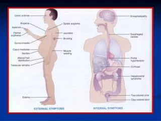
CLINICAL FEATURES History : • Ascites frequently develops as part of the patient's first decompensation of liver disease. It can be associated with other features of liver decompensation such as jaundice or encephalopath.
Risk factors for viral hepatitis, such as ivdu, blood tx, sex, acupuncture, tattoos, ear piercing, and country of origin. • NASH from long-standing obesity, many patients who have been obese will spontaneously lose 50 or even 100 pounds after their liver disease decompensate. • Alcohol intake
Pts with a long history of stable cirrhosis and sudden development of ascites should be suspected of harboring a hepatocellular carcinoma that has caused the decompensation. • 20% of pts with ascites, there is a nonhepatic cause of fluid retention. • Breast, lung, colon, and pancreatic cancers are regularly complicated by ascites. • Malignancy-related ascites frequently is painful, whereas cirrhotic ascites usually is not, unless there is superimposed bacterial peritonitis or alcoholic hepatitis.
A history of heart failure may raise the possibility of cardiac ascites. • Tuberculous peritonitis is usually manifested by fever and abdominal pain, > 50% have underlying alcoholic cirrhosis. • Acute hemorrhagic pancreatitis or hemodialysis. • Fitz-Hugh–Curtis syndrome caused by Chlamydia may cause inflammatory ascites in a sexually active woman. • Pts with ascites and anasarca in the setting of DM suggest nephrotic ascites.
Myxedema and serositis in connective tissue disease may be complicated by ascites. • O/E: • Signs of chronic liver disease • signs of ascites (bulging abdomen,fank dullness,shiffting dullnes and fluid wave). • large veins on the suggests IVC blockage, an immobile mass in the umbilicus (the Sister Mary Joseph nodule) is suggestive of peritoneal carcinomatosis. • Nephrotic syndrome or cardiac failure may have total body edema (anasarca).
Conclusion • The most common cause of ascites is liver cirrhosis and the pathophysiological mechanism is portal HTN leading to systemic vascular changes. • Other pathogenesis include 1-Increased hydrostatic pressure 2-Decreased colloid osmotic pressure 3-Increase in the permeability of peritoneal capillaries 4-Leakage of fluid into the peritoneal cavity 5-miscellaneous