Download
1 / 38
420 likes | 751 Views
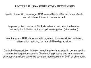
Harini Chandra. Enzymes: Catalytic strategies & regulatory mechanisms.

E N D
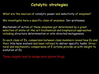
Harini Chandra Enzymes: Catalytic strategies & regulatory mechanisms Binding of a substrate to an enzyme catalytic site brings about conformational changes in the enzyme with multiple weak interactions being formed with the substrate molecule. The amino acid side chains of the enzyme aid in cleavage and formation of bonds thereby providing a variety of mechanisms for catalysis to occur.
Master Layout (Part 1) 1 This animation consists of 4 parts: Part 1 – Covalent catalysis Part 2 – Acid-base catalysis Part 3 – Metal ion catalysis Part 4 – Regulatory strategies Nucleophilic group 2 X X X B A B A 3 Substrate H2O Covalent bond 4 B A Product Product 5 Source: Modified from Biochemistry by A.L.Lehninger et al., 4th edition (ebook)
Definitions of the components:Part 1 – Covalent catalysis 1 1. Covalent catalysis: A catalytic strategy wherein the active site of an enzyme is lined with a reactive group, usually a powerful nucleophile, that undergoes temporary covalent modification during the course of the reaction. A transient covalent bond between the enzyme and substrate facilitates catalysis by providing a pathway with lower activation energy. 2. Substrate: The molecule(s) present at the beginning of a reaction that are modified by enzyme are known as substrate(s). An enzymatic reaction may have one or more substrates depending upon the reaction. 3. Covalent bond: A chemical bond formed between atoms of two reacting species that involves the sharing of outer orbital electrons for bond formation. 4. Nucleophilic group: An electron rich group that readily attacks a positively charged centre to form a chemical bond by donating its electrons. The term nucleophile translates to “nucleus lover”. 5. Product: The molecules produced as a result of an enzymatic reaction are known as the products. A reaction may yield one or more products with the enzyme being regenerated at the end of the reaction. 2 3 4 5
Part 1, Step 1: 1 Nucleophilic group Covalent catalysis H2O Regenerated catalyst 2 A B X 3 Products Substrate Covalent bond 4 Action Description of the action Audio Narration Covalent catalysis involves the formation of a transient covalent bond between the nucleophile present in the enzyme and the substrate molecule. Formation of this bond provides an alternative reaction pathway that has lower activation energy than the uncatalyzed reaction. Several amino acid side chains act as effective nucleophiles that facilitate the reaction. The enzyme is regenerated in its unaltered form at the end of the reaction. As shown in animation. First show ‘A’ and ‘B’ joined by thick line as shown along with ‘X’. ‘X’ must be shown to attack ‘A’ and when this happens, the line between A&B must become a dotted line and a new dotted line must appear between A&X. Next, B must move away and the dotted line must disappear while the dotted line between A&X must become a thick line. Next the blue ‘H2O’ must appear and must pass through the line between A&X. When this happens, the line between A&X must break and they must move apart from each other. 5 Source: Modified from Biochemistry by A.L.Lehninger et al., 4th edition (ebook)
Part 1, Step 2: 1 Example for covalent catalysis - Chymotrypsin O O O O 2 X C C C Ser-195 Ser-195 OH OH R R R Substrate Enzyme H2O X H 3 HO Ser-195 Regenerated enzyme Product 4 Action Description of the action Audio Narration Chymotrypsin is one such enzyme that carries out catalysis by covalent modification. It possess a catalytic triad of histidine, aspartic acid and serine at its active site with the serine at position 195 serving as a highly powerful nucleophile. The reaction between the serine hydroxyl group and the unreactive carbonyl group of the substrate helps in bringing about product formation with regeneration of the enzyme after the reaction. Covalent modification of this serine residue led to irreversible inactivation of the enzyme, which clearly suggested that it performs a vital role in catalysis. First show the ‘enzyme’ and ‘substrate’. Then show the black arrow appearing after which the grey arrow must appear followed by removal of the group ‘XH’ as shown and appearance of the figure after the arrow. The red line shown must then appear. The blue oval marked ‘H2O’ must then appear followed by the black arrow as shown. This must be followed by appearance of the downward grey arrow and the figures shown below it. As shown in animation. 5 Source: Modified from Biochemistry by Lubert Stryer et al., 6th edition (ebook)
Master Layout (Part 2) 1 This animation consists of 4 parts: Part 1 – Covalent catalysis Part 2 – Acid-base catalysis Part 3 – Metal ion catalysis Part 4 – Regulatory strategies R1 R C O 2 N R2 R1 Substrates OH H R O C O Products H N R2 R1 R1 3 H R R C C O O OH O Intermediate N N R2 R2 B Base 4 H H R1 R C O O N H R2 5 BH H
Definitions of the components:Part 2 – Acid-base catalysis 1 1. Acid-base catalysis: Biochemical reactions involving the formation of unstable charged intermediates are often stabilized by transfer of protons to or from the substrate or intermediate. In case of enzyme catalyzed reactions, weak proton donors or acceptors are often present as amino acid side chains at the active site of the enzyme itself. These groups mediate proton transfer reactions which provide rate enhancement of several orders of magnitude. 2. Substrate(s): The molecules present at the beginning of a reaction that are modified by means of the enzyme are known as substrates. An enzymatic reaction may have one or more substrates depending upon the reaction. 3. Enzyme: The biocatalyst responsible for bringing about an increase in the rate of reaction for conversion of substrate to product. 4. Intermediate: Enzymatic reactions proceed through formation of a transition state i.e. an intermediate state in between substrate and product having higher free energy than that of either the substrate or product. The transition state is the least stable species of the reaction pathway due to its high free energy and is therefore the most seldom occupied. 5. Base: An aqueous solution or substance that can accept hydronium ions by donating its free electron pair is known as a base. 6. Product(s): The molecules produced as a result of an enzymatic reaction are known as the products. A reaction may yield one or more products with the enzyme being regenerated at the end of the reaction. 2 3 4 5
Part 2, Step 1: 1 Acid-base catalysis R1 R C O 2 N R2 Substrates OH H 3 R1 R C O OH N Intermediate R2 4 Action Description of the action Audio Narration H First show the two ‘substrates’ on top. The green circle containing ‘OH’ group must move towards the yellow circle containing ‘C’ as shown. The down arrow must then appear to give the figure below. Biochemical reactions involving the formation of unstable charged intermediates are often stabilized by transfer of protons to or from the substrate or intermediate. For non-enzymatic reactions, acid-base catalysis may involve only the hydronium or hydroxyl ions of water, referred to as specific acid-base catalysis. As shown in animation. 5 Source: Modified from Biochemistry by A.L.Lehninger et al., 4th edition (ebook)
Part 2, Step 2: 1 Acid-base catalysis 2 Base 3 R1 R1 R R C C O O OH O N N R2 R2 4 B Action Description of the action Audio Narration H H In many cases, however, water alone does not suffice to catalyze the reaction. In such cases, proton transfer is facilitated by weak organic acids or bases. Organic acids act as proton donors while organic bases can serve as proton acceptors. As shown in animation. Next, show the blue circle ‘base’ appearing which must move towards the green circle containing ’OH’ as depicted. It must then remove the red ‘H’ from it and the figure below must appear. The blue circle must then move away. 5 BH Source: Modified from Biochemistry by A.L.Lehninger et al., 4th edition (ebook)
Part 2, Step 3: 1 Acid-base catalysis 2 3 R1 R C O O N R2 4 B Action Description of the action Audio Narration H R1 In case of enzyme catalyzed reactions, weak proton donors or acceptors are often present as amino acid side chains at the active site of the enzyme itself. The precise positioning of these groups within the active site mediate proton transfer reactions which can provide rate enhancements of several orders of magnitude. Next, the blue circle must move towards the pink circle containing ‘N’. The red ‘H’ must then be transferred to this group as shown in the figure below. The blue circle must then move away. As shown in animation. R C O O 5 N H R2 BH H Source: Modified from Biochemistry by A.L.Lehninger et al., 4th edition (ebook)
Part 2, Step 4: 1 Acid-base catalysis 2 R1 R O C O 3 H N R2 H Products 4 Action Description of the action Audio Narration R1 Acid-base catalysis is a common mechanism of action employed by many enzymes. It is often used in combination with another mechanism such as covalent catalysis. The ease of stabilization of charged intermediates by the amino acid side chains helps in lowering the activation energy for product formation. Show the arrows appearing in the figure above at the positions indicated followed by the downward arrow and the ‘products’ below. As shown in animation. R C O O 5 N H R2 H Source: Modified from Biochemistry by A.L.Lehninger et al., 4th edition (ebook)
Part 2, Step 5: 1 Example for acid-base catalysis - Chymotrypsin Active site – catalytic triad His 57 Ser 195 Asp 102 2 Substrate Chymotrypsin enzyme Oxyanion hole 3 4 Tetrahedral intermediate Action Description of the action Audio Narration (PLEASE RE-DRAW ALL FIGURES.) First show the top left figure appearing with its labels followed by appearance of the arrow and the compound above it. Next the second figure on the right must appear followed by the downward arrow and the figure below it. Chymotrypsin is one such enzyme that employs both covalent catalysis as well as acid-base mechanism. The arrangement of the catalytic triad, consisting of Aspartic acid, Histidine and Serine, at the enzyme’s active site is such that the histidine residue serves as a general base catalyst by polarizing the hydroxyl group of serine. The alkoxide ion thus generated in the serine residue makes it a more powerful nucleophile. As shown in animation. 5 Source: Biochemistry by Lubert Stryer et al., 6th edition (ebook)
Part 2, Step 6: 1 Example for acid-base catalysis - Chymotrypsin Acyl-enzyme intermediate 2 Tetrahedral intermediate Product 1 3 4 Acyl-enzyme intermediate Action Description of the action Audio Narration Following substrate binding and nucleophilic attack of the serine on the carbonyl group, the geometry of the intermediate becomes tetrahedral and the negative charge developed on the carbonyl oxygen gets stabilized through interactions with other side chains of the proteins, in a site known as the oxyanion hole. An internal proton transfer then causes the tetrahedral intermediate to collapse and generate the acyl-enzyme intermediate after which the amine group is released from the active site. (PLEASE RE-DRAW ALL FIGURES.) In continuation with the previous slide. The grey arrow must then appear after ‘tetrahedral intermediate’ followed by the figure on the right. Next the arrow must appear along with the curved arrow showing removal of ‘product 1’ followed by the figure below. As shown in animation. 5 Source: Biochemistry by Lubert Stryer et al., 6th edition (ebook)
Part 2, Step 7: 1 Example for acid-base catalysis - Chymotrypsin Acyl-enzyme intermediate 2 Acyl-enzyme intermediate Oxyanion hole 3 4 Tetrahedral intermediate Action Description of the action Audio Narration Once the amine group leaves the enzyme’s active site, it is replaced by a molecule of water which carries out hydrolysis of the ester group of the acyl-enzyme intermediate. Mechanism for hydrolysis again proceeds via formation of a tetrahedral intermediate with histidine acting as a general acid catalyst and the negative charge on oxygen being stabilized by residues in the oxyanion hole. (PLEASE RE-DRAW ALL FIGURES.) In continuation with the previous slide. The reaction arrow must appear along with entrance of a molecule of ‘H2O’ as shown. This is followed by appearance of the figure on the right after which the downward arrow must appear along with the figure below. As shown in animation. 5 Source: Biochemistry by Lubert Stryer et al., 6th edition (ebook)
Part 2, Step 8: 1 Example for acid-base catalysis - Chymotrypsin 2 Tetrahedral intermediate Product 2 3 4 Regenerated enzyme Action Audio Narration Description of the action The tetrahedral intermediate then breaks down to liberate the second product in the form of a carboxylic acid along with regeneration of the enzyme which is then ready for another round of catalysis. Internal proton transfers between amino acid side chains therefore play a vital role in acid-base catalysis by enzymes. (PLEASE RE-DRAW ALL FIGURES.) In continuation with the previous slide. The arrow must then appear to show the figure on the right top followed by the downward arrow and removal of the compound shown as ‘product 2’ followed by the appearance of the figure below. As shown in animation. 5 Source: Biochemistry by Lubert Stryer et al., 6th edition (ebook)
Master Layout (Part 3) 1 This animation consists of 4 parts: Part 1 – Covalent catalysis Part 2 – Acid-base catalysis Part 3 – Metal ion catalysis Part 4 – Regulatory strategies R1 R1 R1 H 2 M2+ M2+ C C C O O O H Metal ion Substrate O O R2 R2 R2 H H 3 H2O H2O 4 M2+ M2+ O O H H Regenerated catalyst Product Intermediate 5 Source: Modified from Biochemistry by Lubert Stryer et al., 6th edition (ebook)
Definitions of the components:Part 3 – Metal ion catalysis 1 1. Metal-ion catalysis: Metal ions, either present in solution or bound to the enzyme itself, facilitate catalysis by forming favorable interactions that orient the substrate and enzyme in suitable positions for transition state, and subsequently, product formation. These interactions help in stabilizing the intermediates formed, thereby lowering the activation energy required for reaction to occur. 2. Substrate(s): The molecules present at the beginning of a reaction that are modified by means of the enzyme are known as substrates. An enzymatic reaction may have one or more substrates depending upon the reaction. 3. Enzyme: The biocatalyst responsible for bringing about an increase in the rate of reaction for conversion of substrate to product. 4. Intermediate: Enzymatic reactions proceed through formation of a transition state i.e. an intermediate state in between substrate and product having higher free energy than that of either the substrate or product. The transition state is the least stable species of the reaction pathway due to its high free energy and is therefore the most seldom occupied. 5. Metal ion: Metals usually present in their divalent state (i.e. +2 charge) facilitate catalysis. One or more metal ion has been found to be required for catalysis of nearly one third of all enzymatic reactions. One of the most commonly found metal ions in enzymatic mechanisms is Zn2+. 6. Product(s): The molecules produced as a result of an enzymatic reaction are known as the products. A reaction may yield one or more products with the enzyme being regenerated at the end of the reaction. 2 3 4 5
Part 3, Step 1: 1 Metal ion catalysis H M2+ O Metal ion H 2 H M2+ Water 3 O Strong nucleophile generated H 4 Action Description of the action Audio Narration First show the purple oval along with the ‘water’ figure. The purple circle must then approach the blue circle of water. When this happens, the red line must appear between the two and one green ‘H’ must leave in the form of ‘H+’ as depicted to give the figure on the right. As shown in animation. Metal ions, either present in solution or bound to the enzyme itself, facilitate catalysis by forming favorable interactions between enzyme and substrate or in the transition state. Metal ions in the active site of the enzyme typically react with a water molecule and activate it by facilitating generation of a strong nucleophile in the form of a hydroxide ion. 5 Source: Modified from Biochemistry by Lubert Stryer et al., 6th edition (ebook)
Part 3, Step 2: 1 Metal ion catalysis 2 R1 R1 M2+ C C O O 3 Strong nucleophile generated O R2 R2 Intermediate Substrate H 4 Action Description of the action Audio Narration As shown in animation. The nucleophilic alkoxide ion attacks the unreactive carbonyl group to form a tetrahedral intermediate in which the charges are stabilized by the metal ion. These favorable interactions help in orienting the substrate and enzyme in suitable positions for transition state, and subsequently, product formation. M2+ In continuation with previous slide. Show the ‘substrate’ molecule appearing after which the black arrows must appear as shown. The grey arrow must then appear along with the figure on the right. O H 5 Source: Modified from Biochemistry by Lubert Stryer et al., 6th edition (ebook)
Part 3, Step 3: 1 Metal ion catalysis H2O 2 R1 R1 C C O O 3 R2 R2 Regenerated catalyst Product Intermediate H2O 4 Action Description of the action Audio Narration M2+ In continuation with previous slide. Show the blue ‘H2O’ appearing which must move towards the violet oval as shown. This is followed by appearance of the small black arrow after which the grey arrow and the figures on the right must appear as depicted. As shown in animation. M2+ Hydrolysis of the stabilized intermediate leads to product formation along with liberation of the regenerated metal catalyst. Thus by lowering the activation energy of the transition state, metal ions facilitate enzyme catalyzed reactions. Almost one third of known enzymes have been found to require one or more metal ions for their activity. O O H H 5 Source: Modified from Biochemistry by Lubert Stryer et al., 6th edition (ebook)
Part 3, Step 4: 1 Example for metal ion catalysis – Carbonic anhydrase H His 119 His 119 2 H Zn2+ Zn2+ His 96 His 96 O His 94 His 94 H 3 O Water Active site H Strong nucleophile generated Carbonic anhydrase 4 Action Description of the action Audio Narration First show appearance of ‘carbonic anhydrase’, ‘active site’ and ‘water’. This is followed by appearance of the black arrow mark as shown after which the grey arrows, the figure on the right and the green ‘H+’ must appear. Carbonic anhydrase is an enzyme responsible for hydration and dehydration reactions of carbon dioxide and bicarbonate respectively and has been found to have a divalent zinc ion associated with its activity. The zinc ion in its active site is bound to the imidazole rings of three histidine residues as well as to a molecule of water. This binding to water facilitates formation of the hydroxide nucelophile with concomitant release of a proton. As shown in animation. 5 Source: Modified from Biochemistry by Lubert Stryer et al., 6th edition (ebook)
Part 3, Step 5: 1 Example for metal ion catalysis – Carbonic anhydrase O O His 119 His 119 C C 2 Zn2+ Zn2+ His 96 His 96 O O Substrate His 94 His 94 3 O O H H Electrostatic stabilization forces 4 Intermediate Action Audio Narration Description of the action The generated hydroxide ion at the active site then attacks the carbon dioxide substrate, converting it into a bicarbonate ion. The negative charge generated on the oxygen atom is stabilized by interactions with the zinc ion. In continuation with previous slide. The ‘substrate’ should then appear after which the black arrows must be shown as depicted. This is followed by the grey downward arrow and the figure show below. The dotted red line must appear in the end after the figure is shown as depicted in animation. As shown in animation. 5 Source: Modified from Biochemistry by Lubert Stryer et al., 6th edition (ebook)
Part 3, Step 6: 1 Example for metal ion catalysis – Carbonic anhydrase O O His 119 His 119 C C 2 H H Zn2+ Zn2+ Regenerated enzyme His 96 His 96 O O O O His 94 His 94 H H 3 O H O H Product (bicarbonate) 4 Action Audio Narration Description of the action In continuation with previous slide. Another ‘water’ molecule shown above must then appear and approach the brown oval. The grey arrow must then appear followed by the figures shown on the right. Binding of another molecule of water to the zinc ion at the active site of the enzyme leads to release of the bicarbonate ion and regeneration of the enzyme molecule ready for another round of catalysis. Several pH related studies have provided substantial proof for this mechanism. As shown in animation. 5 Source: Modified from Biochemistry by Lubert Stryer et al., 6th edition (ebook)
Master Layout (Part 4) 1 This animation consists of 4 parts: Part 1 – Covalent catalysis Part 2 – Acid-base catalysis Part 3 – Metal ion catalysis Part 4 – Regulatory strategies Enzyme regulatory strategies 2 3 Reversible covalent modification Allosteric/ feedback inhibition Isozyme forms Proteolytic activation 4 5 Source: Modified from Biochemistry by Lubert Stryer et al., 6th edition (ebook)
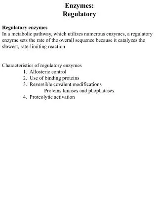
Definitions of the components:Part 4 – Regulatory strategies 1 1. Allosteric/feedback inhibition: Enzymes that contain distinct catalytic and regulatory sites are said to be allosteric in nature. Binding of regulatory molecules at the regulatory sites triggers off a series of conformational changes that ultimately reach the active site and prevents binding of substrate molecule thereby regulating catalysis. In many pathways, the reaction that is regulated involves the enzyme catalyzing the first step of the pathway so that all further reactions taking place after the first also automatically get regulated due to lack of substrate availability. When this enzyme is regulated by the final product of the reaction pathway, it is known as feedback inhibition. Most allosteric enzymes are subject to feedback control, a popular example being aspartate transcarbamoylase. 2. Isozyme forms: Isoenzymes are homologous enzymes within a single organism that differ slightly in their structure but catalyze the same reaction. They are mostly expressed in different tissues and have differing kinetic parameters such as substrate affinity (Km) and maximum velocity (Vmax). These differing properties allow the isozyme forms to be differentially regulated at varying points of time. Example: Lactate dehydrogenase exists in different forms in heart and muscle tissue. The heart isozyme has a higher affinity for substrates and is allosterically inhibited by high levels of pyruvate while the muscle enzyme is not. 3. Reversible covalent modification: Several covalent modifications such as phosphorylation, uridylylation, methylation etc. significantly alter the catalytic activity of enzymes. Covalently modified enzymes may either have increased or decreased activity for the reaction it catalyzes. The enzymes responsible for covalent modification of other enzymes are usually regulated by further control mechanisms. Example: Enzymes of glycogen metabolism such as glycogen phosphorylase are regulated by covalent modification. 2 3 4 5
Definitions of the components:Part 4 – Regulatory strategies 1 4. Proteolytic activation: Many enzymes exist in their inactive forms, known as zymogens, and need to be activated by hydrolysis of one of more peptide bonds. This removal of certain regulatory residues irreversibly converts the enzyme into its active form. The enzyme is then degraded after completion of catalysis. Example: Digestive enzymes such as trypsin, chymotrypsin as well as blood clotting factors are regulated by mechanism of proteolytic activation. 2 3 4 5
Part 4, Step 1: 1 Enzyme regulatory strategies 2 3 Allosteric/ feedback inhibition Reversible covalent modification Isozyme forms Proteolytic activation 4 Action Description of the action Audio Narration (Please redraw figures.) The top heading must be shown followed by the four arrows and the headings in the circles below. The user should be allowed to click on any of the headings in the circles to view the description provided in the previous two slides. Another ‘proceed’ tab must be present on the right bottom and when user clicks on that, the text in the blue circle and yellow circle must be highlighted after which it must move to the next slide. Activity of all enzymes must be regulated to ensure that they function only to the desired extent at the appropriate locations within an organism. Common mechanisms of regulation include allosteric or feedback inhibition, control of isozyme forms, reversible covalent modification and proteolytic activation. <If user clicks on any of these four tabs, the description as given in previous two slides must be narrated.> As shown in animation. 5 Source: Modified from Biochemistry by Lubert Stryer et al., 6th edition (ebook)
Part 4, Step 2: 1 Allosteric/feedback inhibition Substrate B Substrate C Substrate D 2 Substrate A Enzyme D Enzyme B Enzyme C 3 Substrate E Enzyme A Allosteric enzymes DO NOT obey Michaelis-Menten kinetics. 4 Action Description of the action Audio Narration Allosteric enzymes, which possess distinct regulatory and catalytic sites, are often found as the first enzyme of a reaction pathway. Regulation of the first enzyme of a pathway by the final product of the pathway is known as feedback inhibition. Binding of the regulator molecule to the regulatory site of the enzyme triggers a series of conformational changes that are ultimately transmitted to the active site where substrate binding is then inhibited. It has been observed that allosteric enzymes do not obey regular Michaelis-Menten kinetics. As shown in animation. First show blue ‘enzyme A’ along with green ‘substrate A’. Green substrate must bind in the pocket as shown after which its shape must change to a brown oval. This oval must leave the pocket after which the sequence of arrows and the other shapes must appear as depicted in animation. When ‘Substrate E’ appears, it must move towards ‘enzyme A’ and bind in the other pocket as depicted. When this happens, the pocket on top must change shape as shown and the green substrate should not be able to bind to it. Then red crosses must appear across all the other reaction arrows as well. 5 Source: Modified from Biochemistry by Lubert Stryer et al., 6th edition (ebook)
Part 4, Step 3: 1 Example for allosteric/feedback inhibition – Aspartate Transcarbamoylase Catalytic subunits Regulatory subunits 2 Inhibitor Substrate CTP CTP CTP CTP CTP PALA PALA PALA Enzyme 3 Stabilized R state 4 Stabilized T state Action Description of the action Audio Narration Aspartate transcarbamoylase, which catalyzes the first step of pyrimidine biosynthesis, is an allosteric enzyme having distinct regulatory and catalytic subunits. Binding of substrate to the catalytic subunits induces conformational changes that stabilize the relaxed state or R state of the enzyme, thereby facilitating the enzymatic reaction. The inhibitor for this enzyme is CTP which is the final product of the pathway. Binding of inhibitor to the regulatory subunits stabilizes the tense state or T state of the enzyme thereby preventing the reaction from taking place. As shown in animation. PLEASE REDRAW ALL FIGURES. First show the figure on top with its labels. Next show the left arrow appearing with the violet circles binding to the red circles as shown. Next show the right arrow appearing with blue circles binding to the yellow regions as shown. 5 Source: Biochemistry by Lubert Stryer et al., 6th edition (ebook)
Part 4, Step 4: 1 Isozyme forms Different regulatory molecules Substrate Substrate Different regulatory site 2 3 Tissue A – isozyme A Tissue B – isozyme B Similar catalytic site Same reaction catalyzed – different kinetic parameters such as Vmax, Km. Product Product 4 Action Description of the action Audio Narration First show the green & brown ‘isozymes A&B’ appearing. The arrows with labels must appear, flash a couple of times and then disappear. Next, the ‘substrate’ must appear, enter the ‘catalytic site’ as shown where it must change shape and then leave as the ‘product’. This must take place simultaneously for both A&B. The reaction taking place in B must be faster than A as depicted in animation. Once this happens, the text message shown below in red must appear. Next the violet pentagon must bind to the ‘regulatory site’ on top in B but not in A as depicted. This is followed by text message on top. Isoenzymes are homologous enzymes within a single organism that differ slightly in their amino acid sequence but catalyze the same reaction. These enzymes are mostly expressed in different tissues and have differing kinetic parameters such as substrate affinity (Km) and maximum velocity (Vmax). They may also have different regulator molecules that allow them to be differentially expressed and regulated depending on the requirement for that particular tissue. As shown in animation. 5 Source: Modified from Biochemistry by Lubert Stryer et al., 6th edition (ebook)
Part 4, Step 5: 1 Example for isozyme regulation – Lactate dehydrogenase MUSCLE ISOZYME – M4 HEART ISOZYME – H4 Lactate 2 High affinity for substrate Low affinity for substrate 3 LDH LDH Not inhibited by pyruvate Inhibited by pyruvate Pyruvate 4 Action Description of the action Audio Narration As shown in animation. PLEASE REDRAW ALL FIGURES. First show the figure of heart and muscle along with the ‘LDH’ enzyme. Next show the reaction in the centre followed by the text ‘high affinity…’. Next show the blue pentagon binding to the pocket on the left but being unable to bind on the right followed by the text below as depicted. Lactate dehydrogenase is an enzyme involved in anaerobic glucose metabolism that is present as two isozyme forms in human beings. The tetrameric heart enzyme , which requires an aerobic environment to function, has higher affinity for its substrate than the muscle enzyme. Despite having 75% sequence homology, they also differ in that high levels of pyruvate allosterically inhibit the heart enzyme but not the muscle form. 5 Source: Modified from Biochemistry by Lubert Stryer et al., 6th edition (ebook)
Part 4, Step 6: 1 Reversible covalent modification Pi ATP H2O ADP Substrate P Substrate 2 Kinase Enzyme Enzyme Phosphatase More Active Less Active 3 Product Product 4 Action Description of the action Audio Narration Reversible covalent modification is another commonly employed enzyme regulatory strategy. The most widely observed modification is phosphorylation, which is carried out by various enzyme kinases with the help of ATP as a phosphoryl donor. Some enzymes are more active in their phosphorylated forms while others are less active in this form. Dephosphorylation is carried out by the phosphatase enzyme. Enzymes involved in glycogen metabolism are regulated by reversible phosphorylation. As shown in animation. First show reaction on the left appearing along with the ‘less active enzyme’. A red cross must appear over the reaction arrow. The curved arrows towards the right must then appear followed by the ‘more active enzyme’ on the right. When this appears, the reaction must take place as shown on right. Finally the curved arrows towards the left must appear as depicted. 5 Source: Modified from Biochemistry by A.L.Lehninger et al., 4th edition (ebook)
Part 4, Step 7: 1 Reversible covalent modification 2 3 4 Action Description of the action Audio Narration Apart from phosphorylation, which most commonly takes place at serine, threonine and tyrosine residues, other reversible covalent modifications include adenylylation, uridylylation, methylation and ADP-ribosylation which modify different amino acid residues of the proteins. As shown in animation. The rows of the table must appear one at a time. 5 Source: Modified from Biochemistry by A.L.Lehninger et al., 4th edition (ebook)
Part 4, Step 8: 1 Proteolytic activation Enzyme inactive form (zymogen) Enzyme active form 2 Protease 3 Substrate Product 4 Action Description of the action Audio Narration As shown in animation. First show the reaction coming in as depicted followed by the ‘inactive’ enzyme’ appearing on the arrow followed by the red cross. Next show the pie shaped object ‘protease’ entering which must cleave the red portion and remove it. The red cross must then disappear and the label for enzyme must change as shown. Several enzymes exist in their inactive forms, known as zymogens, where they do not possess any catalytic activity. In order to become active, they need to be activated by hydrolysis of one of more peptide bonds by various protases. This removal of certain regulatory residues irreversibly converts the enzyme into its active form. Unlike reversible modification, the enzyme is degraded after completion of catalysis. 5 Source: Modified from Biochemistry by Lubert Stryer et al., 6th edition (ebook)
Part 4, Step 9: 1 Example for proteolytic activation – Chymotrypsin Chymotrypsinogen (inactive) 1 245 Substrate 2 Product 1 Trypsin 245 15 16 Pi-chymotrypsin 3 Pi-chymotrypsin 1 13 16 149 245 146 Substrate Product B-chain A-chain C-chain 4 a-chymotrypsin (active) Action Description of the action Audio Narration Several digestive enzymes as well as clotting factors are regulated by proteolytic activation. Chymotrypsin, a digestive enzyme that hydrolyzes proteins in the small intestine, exists in its zymogen form within membrane bound granules after synthesis in the acinar cells of the pancreas. The proteolytic enzyme trypsin converts it into its fully active form by cleavage of a peptide bond between arginine at position 15 and isoleucine at position 16. The resulting enzyme known as pi-chymotrypsin is acted upon by other such molecules to yield the completely active and stable alpha-chymotrypsin which consists of three chains linked by inter-chain disuphide bonds. As shown in animation. First show the ‘inactive chymotrypsinogen’ above followed by appearance of the reaction on the left with the red cross on it. Next show the ‘trypsin’ which must cleave the blue rod as shown to give ‘pi-chymotrypsin’. The green ‘trypsin’ must again cleave the blue chain to give the figure below of alpha-chymotrypsin followed by the reaction sequence on the right as depicted. 5 Source: Modified from Biochemistry by Lubert Stryer et al., 6th edition (ebook)
Interactivity option 1:Step No:1 1 Match the following enzymes with their appropriate regulatory strategies. Enzyme name Regulatory mechanism 2 Glycogen phosphorylase 1. Isozyme forms A. Aspartatetranscarbamoylase 2. Covalent modification B. 3 Chymotrypsin 3. Allosteric inhibition C. Lactate dehydrogenase 4. Proteolytic activation D. 4 Results Interacativity Type Options User must drag & drop the A, B, C, D provided in the left column into the dotted boxes present in the right column. Every time the user drags and matches it correctly, that box must then turn green. If user gets it wrong, then it must move back automatically into the left column. Correct answers are 1-D, 2-A, 3-B and 4-C. User must be allowed to drag the A, B, C, D into the dotted boxes shown on the right. Match the following. 5
Questionnaire 1 1. Which of the following amino acid residues is not part of the catalytic triad of the active site of chymotrypsin? Answers: a) His b) Asp c) Ser d) Pro 2. Which metal ion bound to the enzyme carbonic anhydrase is responsible for its activity? Answers:a) Zn2+ b) Fe2+ c) Mg2+ d) Mn2+ 3. The attachment of a metal ion to water generates which of the following molecules? Answers:a) Strong electrophileb)Strong nucleophilec) Weak electrophile d) Weak nucleophile 4. S-adenosyl methionine is the donor group for which type of reversible covalent modification? Answers: a) Phosphorylation b) Adenylylation c) Methylation d) UDP-ribosylation 2 3 4 5
Links for further reading Books: Biochemistry by Stryer et al., 5th & 6th edition Biochemistry by A.L.Lehninger et al., 4th edition Biochemistry by Voet & Voet, 3rd edition Fundamentals of Enzymology by Price & Stevens, 3rd edition