Download
1 / 44
530 likes | 849 Views
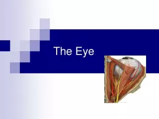
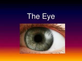
The Eye. by: Elora Zavala and Hallesha Williams. General Facts of the Eye. Purpose: to help see what is around you. About 1 inch in diameter Made up of 3 layers Outermost Cornea Sclera Middle Layer Choroid Ciliary Body Iris Innermost Retina. Cornea.

E N D
The Eye by: Elora Zavala and Hallesha Williams
General Facts of the Eye • Purpose: to help see what is around you. • About 1 inch in diameter • Made up of 3 layers • Outermost • Cornea • Sclera • Middle Layer • Choroid • Ciliary Body • Iris • Innermost • Retina
Cornea • Clear, dome shaped surface that covers the iris, pupil, and anterior chamber. • Most powerful lens that has no blood vessels • Contains Five Layers • Epithelium • Anterior Elastic Lamina • Substantia Propria • Posterior Elastic Lamina • Corneal Endothemlium
Sclera • White protective part of the eye • Has Four layers • Episclera • Stroma • Lamina Fusca • Endothelium
Choroid • Layer of blood vessels between the retina and sclera • Supplies blood to retina
Ciliary Body • Where aqueous humor is produced • Attached to lens by zonules • Also controls focus by changing the shape of the lens.
Iris • Colored part of the eye that is surrounded by sclera • Ring of muscle fibers located behind cornea and in front of lens. Consists of two layers • Pigmented Epithelial Cells • Pigmented Fibrovascular Tissue (Stroma) • Connected to a sphincter muscle that controls dilation and contraction of pupil. • Helps protect the sensitive retina
Retina • Thin nerve membrane that detects light entering the eye
Retinal Blood Vessels • Supply blood to the retina and are visible to the eye • Located in the choroid just beneath retina
Retinal Pigment Epithelium • Layer of cells between the retina and choroid • Melanin in the RPE gets rid of waste products
Pupil • The opening in the iris. • Pupil size is seen by the iris contraction or dilations.
Crystalline Lens • Located behind the cornea, normally clear • Light passes through pupil to lens • Small muscles attached to lens
Vitreous Humor • Jelly like, thick liquid that fills the eye to help maintain its shape • Located between the the lens and retina • Drains back into the blood through canals of schlemm
Canals of Schlemm • Located around the perimeter of the iris • Allows vitreous gel (or aqueous humor) to drain back into blood stream
Vitreous Cavity • The space between the lens and retina filled with gel.
Visual Fields • Retina of each eye has two sections • Nasal Retina (Yellow) • Temporal Retina (Green)
Optic Nerve • The nerve at the back of the eye that carries visual information from the eye to the brain. • Blind spot comes from the optic disc located near the optic nerve.
Macula • Near the center of the retina at the back of the eyeball. • This part of the eye gives us our 20/20 vision.
Fovea • Indentation in the center of the macula • Is responsible for our highest visual acuity • The center of our central vision
Uvea • Middle vascular layer of the eye • Three Parts • Iris • Ciliary Body • Choroid
Zonules • Hundreds of string like fibers that hold in position • Enable it to change shape for near or distant vision
Fluid Chambers of the Eye • Anterior Chamber • Space between cornea and iris filled with aqueous humor. • Aqueous Humor • Fluid produced in the eye • Posterior Chamber • Space between iris and lens filled with aqueous humor.
Muscles of the Eye • Ciliary Muscle • Changes the shape of the lens • Flattens lens for distance vision • Contracts for closer vision • Produces aqueous humor.
Muscles of Eye cont. Muscles located OUTSIDE of the eye • Superior Rectus - rotates the eye upwards • Inferior Rectus - rotates the eye downwards • Medial Rectus - rotates the eye towards the nose • Lateral Rectus - rotates the eye towards the ear • Superior Oblique - aids in upward movement • Inferior Oblique - aids in downward movement
Accessory Organs • Eyelashes/Eyebrows • Specialized hairs that protect the eye for dust and insects • Conjunctiva • Thin, clear membrane located on the rim of bottom inner eyelids and covering front of the eye. • Cells produce mucous to help lubricate the eye • Inflammation is conjunctivitis, commonly known as pink eye.
Accessory Organs: cont. • Eye Socket • Cone shaped bone cavity that protects the eye • Padded with fatty tissue • Eyelids • Protects and lubricates the eye • Lines the inner edge of the eyelid.
Accessory Organs: cont. • Tarsus • Supports the eyelid skin • Gives the lid its- • Strength • Shape • Place for muscles to attach
Accessory Organs: cont • Lacrimal Sac • Drains tears and other debris from eye • Lacrimal Glands • Releases tears and other protective fluid onto the surface of the eye • Keeps cornea from being dehydrated
Accessory Organs: cont. • Visual Cortex • Part of the brain that processes and combines visual information both eyes and converts it into sight • Visual Axis • Imaginary line drawn from the center of the pupil to the center of fovea. • Fixation Point comes from the visual axis
Accessory Organs: cont. • Optic Chiasm • First part of the brain to receive visual input • Each eye takes a slightly different picture of the world
Neurons • Ganglion Cells • Located near the inner surface of the retina of the eye. • Receives visual information from photoreceptors via 2 intermediate neuron types: Bipolar and Amacrine cells. • Amacrine Cells • Considered a supplement to the action of horizontal cells • Horizontal Cells • Allows eyes to adjust to the bright and dim lights around them • Bipolar Cells • Transmit signals from the photoreceptors to the ganglion cells
Neurons: cont. • Pigment Epithelium • Shields the retina from excess incoming light • Rods/Cons • AKA Photoreceptors • Found in the retina. • Convert light into signals that can stimulate biological processes.
Cranial Nerves (involved with sight) • Optic (II) • Sensory • Sensory fibers transmit impulses associated with sense of vision • Oculomotor (III) • Motor fibers transmit impulses to muscles that raise eyelids, move the eyes, adjust amount of light that enters the eye, and focuses the lenses
Cranial Nerves cont. • Trochlear (IV) • Motor fibers transmit impulses to muscles that move the eyes • Opthalmic • Sensory fibers transmit impulses from the surfaces of the eyes, tear glands, upper eyelids, etc. • Abducens (VI) • Motor fibers transmit impulses to the muscles that move the eys
Visual Receptors • Visual receptor cells are a layer of rods and cones (the photoreceptor cells I mentioned earlier) that aid in visual inside of retina • Each rod or cone contains a pigment that absorbs a certain type of wavelength better than others
Refraction • Makes image formation possible • When light travels through the lens it’s path is bent or refracted. • The eye itself, sees an image upside down but the signal to the brain flips it right side up.
Pigments Iodopsin Rhodopsin The pigment sensitive to red light in the retinal rods of the eyes, consisting of opsin and retinene. Also called visual purple • a violet light-sensitive pigment in the cones of the retina of the eye that is responsible for color vision
Dark VS Light Vision Dark Light Cons are responsible Eyes can see best in Responsible for color and fine detail. • Rods are responsible • Only can tell between black and white shade. • Provides enhanced sensitivity
Convergent VS Divergent Waves Convergent Waves Divergent Waves Eyes begin to look outward AKA Lazy Eye • Eyes begin to look inward • AKA Crossed Eyed
Stereoscopic Vision • AKA Binocular Vision • Provides information to the brain to find the depth of the visual scene which is also known as 3D sight
EYE SEE YOU! http://www.youtube.com/watch?v=gvozcv8pS3c
Works Citied http://www.aoa.org/x5352.xml http://www.allaboutvision.com/eye-exam/refraction.htm http://hyperphysics.phy-astr.gsu.edu/hbase/vision/rfreye.html http://hubel.med.harvard.edu/book/b41.htm http://faculty.stcc.edu/AandP/AP/AP2pages/Units14to17/unit15/retina.htm http://www.99main.com/~charlief/Blindness.htm http://www.webmd.com/eye-health/structures-of-the-eye http://www.yorku.ca/eye/blndspot.htm http://www.ophthobook.com/chapters/anatomy http://www.stlukeseye.com/anatomy/ciliary.html http://www.gopetsamerica.com/anatomy/iodopsin.aspx http://www.vetmed.vt.edu/education/curriculum/vm8054/eye/rhodopsn.htm http://www.shreveporteyeclinic.com/humaneye_anatomy_details.asp http://www.ncbi.nlm.nih.gov/pubmed/1492139