Download
1 / 43
430 likes | 659 Views
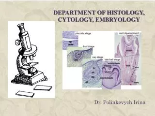
Female Embryology, Anatomy and Histology. A Gari MD. 1-Embryology. Gonads. Internal Genitalia. External Genitalia. A-Embryology (Gonads). Starts @ 4 weeks. Thickened peritoneal (coelomic) membrane (genital ridge) Y chromosome can be identified by 7 w.

E N D
Female Embryology, Anatomy and Histology A Gari MD.
1-Embryology • Gonads. • Internal Genitalia. • External Genitalia.
A-Embryology (Gonads) • Starts @ 4 weeks. • Thickened peritoneal (coelomic) membrane (genital ridge) • Y chromosome can be identified by 7 w. • First follicles is seen @ about 20 weeks.
B-Embryology (Internal Genitalia) • Paramesonephric duct (mullerian) vs. • Mesonephric duct (wollfian).
Upper vagina, cervix, uterus and tubes are formed from the paramesonephric ducts. • Absence of Y chromosome is the key factor. • If present… MIF • If not…regression of wollfian ducts (10-16w)
C-Embryology (External Genitalia) • By 7th week M&F appearance are the same. • Genital tubercle and urogenital membrane (endo/ecto dermal cells) *Genital folds (laterally)… Labia Majora *Urogenital folds (medially)… Labia Minora, Prepuce and Clitoris.
2 - Female Anatomy • Abdominal cavity:
Female Anatomy • Abdominal wall (gross anatomy)
The Uterus thick walled pear shaped muscular organ. Usually anteverted anteflexed, approximately 7.5 cm. Long, 5cm broad. The uterus is located inside the pelvis immediately dorsal to the urinary bladder and ventral to the rectum. The myometrium is 3 lyers L-O-L The Internal Organs
Vagina: • H shape with rugea. • No glands • 8-12 cm in length. • Supplied with (vaginal art.). • Lymphatics varies according to the segment.
Cervix: • 3-4cm in length X 8 mm • Uterine / Cx ratio varies with age. • Columnar epith. • Blood supply (3-9 o'clock) • Lymphatics:
Tubes: • 10-14-cm • 4 segments • Interstitial 2 cm. • Isthmus 4 cm • Ampullary 4-6-cm • Infundibulum 20-25 finger projections. • Inner circular and outer longitudinal muscles. • Blood supply: uterine & ovarian art.
Ovaries: • 1.5x2.5x4 cm • Rests on the ovarian fossa. • IP & Ovarian ligaments: • Blood supply: Art Vs Venous.
Bartholin’s glands. • Skene’s glands.
3- Histology of the Female Genital tract • Vulva: -L Majora: Cornified squamous epithelium. -L Minora: Less Cornified w/ no hair follicle, erectile CT
vagina A - Mucosa - The stratified squamous epithelium • deep stratum basalis • intermediate stratum spinosum • superficial layers of flat eosinophilic cells, contain keratin but (do not horny layer). • typical erectile tissue. B - Muscularis - Inner circular and outer longitudinal layers of smooth muscle are present. - Inferiorly, the striated, voluntary bulbospongiosus muscle forms a sphincter around the vagina. C - Adventitia - Bordering the muscularis, contains many elastic fibres.
Cervix • Is made up of epithelium and underlying stroma. • The stroma contains an admixture of smooth muscle and fibrous and elastic tissues. • The ectocervix is lined by nonkeratinizing stratified squamous epithelium. • The endocervix is covered by mucin-secreting, simple columnar epithelium, • The cells seen in a Pap smear: cells of the ectocervical and endo-cervix. • The border between the stratified squamous epithelium of the ectocervix and the columnar epithelium of the endocervix is called the squamocolumnar junction (SCJ). • Original SCJ: is the site at which the neonatal squamous epithelium of the ectocervix meets the endocervical columnar epithelium at birth. • New, functional or physiologic SCJ: newly formed SCJ as a result of the dynamic remodeling that takes place during the life of the female.
Uterus A - The Myometrium • The muscle fibres of the uterus form layers with preferred orientations of fibres (actually 3-4), but this is very difficult to see in most preparations. • L.O.L B - The Endometrium • consists of a simple columnar epithelium (ciliated cells and secretory cells) and stroma • The mucosa forms many simple uterine glands. • The endometrium is subject to cyclic changes that result in menstruation. • divided into two 1- basalis and 2- functionalis. • The basalis is not sloughed off during menstruation but functions as a regenerative zone for the functionalis after its rejection. • The functionalis is sloughed off during every menstruation. • These cyclic changes are divided into proliferative (or follicular), secretory (or luteal), and menstrual. C - Serosa
Tubes (Oviducts) A - The mucosa • Is formed by a ciliated and secretory epithelium. • The number of ciliated cells and secretory cells varies along the oviduct. B - The muscularis • inner circular muscle layer. • outer longitudinal layer. • inner longitudinal layer is present in the isthmus and the intramural part of the oviduct. • C – The Serosa