Download
1 / 53
530 likes | 695 Views
Membrane Structure and Function. Membrane Function. Membranes organize the chemical activities of cells. The outer plasma membrane forms a boundary between a living cell and its surroundings Exhibits selective permeability Controls traffic of molecules in and out. Membrane Function.

E N D
Membrane Function • Membranes organize the chemical activities of cells. • The outer plasma membrane • forms a boundary between a living cell and its surroundings • Exhibits selective permeability • Controls traffic of molecules in and out
Membrane Function • Internal membranes provide structural order for metabolism • Form the cell's organelles • Compartmentalize chemical reactions
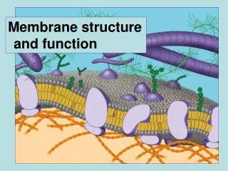
Fluid Mosaic Model of the PM • A membrane is a mosaic • Proteins and other molecules are embedded in a framework of phospholipids • A membrane is fluid • Most protein and phospholipid molecules can move laterally
Membrane Structure Phospholipid Phospholipids are the major structural component of membranes.
Membrane Structure All membranes are phospholipid bilayers with embedded proteins. Phospholipid Bilayer Label the: Hydrophilic heads Hydrophobic tails
Embedded in the bilayer are proteins • Most of the membrane’s functions are accomplished by the embedded proteins. • Integral proteins span the membrane • Peripheral proteins are on one side or the other of the membrane
Plasma Membrane Components • Glycoproteins and glycolipids are proteins/lipids with short chain carbohydrates attached on the extracellular side of the membrane.
Carbohydrate of glycoprotein Glycoprotein Fig. 5-1a Glycolipid Integrin Phospholipid Microfilaments of cytoskeleton Cholesterol
Types of Membrane Proteins • Cell-cell recognition proteins • Integrins • Intercellular junction proteins • Enzymes • Signal transduction proteins • Aka - Receptor proteins • Transport proteins • Passive and active
Cell-cell recognition proteins - identify type of cell and identify a cell as “self” versus foreign • Most are glycoproteins • Carbohydrate chains vary between species, individuals, and even between cell types in a given individual. • Glycolipids also play a role in cell recognition
Integrins are a type of integral protein • The cytoskeleton attaches to integrins on the cytoplasmic side of the membrane • Integrins strengthen the membrane • Intercellular junction proteins - help like cells stick together to form tissues
Many membrane proteins are enzymes • This is especially important on the membranes of organelles.
Signal transduction (receptor) proteinsbind hormones and other substances on the outside of the cell. • Binding triggers a change inside the cell. • Called signal transduction • Example: The binding of insulin to insulin receptors causes the cell to put glucose transport proteins into the membrane.
Messenger molecule Fig. 5-1c Receptor Activated molecule
Transport Proteins • Passive Transport Proteins • allow water soluble substances (small polar molecules and ions) to pass through the membrane without any energy cost • Active Transport Proteins • The cell expends energy to transport water soluble substances against their concentration gradient
Transport of Substances Across the Plasma Membrane (PM) • Passive Transport • (Simple) Diffusion (5.3) • Facilitated diffusion (5.6) • Osmosis (5.4, 5.5) • Active Transport (5.8) • Bulk Flow (5.9) • Endocytosis • Exocytosis
Passive Transport • In passive transport substances cross the membrane by diffusion • Diffusion - net movement of substances from an area of high concentration to low concentration • no energy required
Factors Affecting Diffusion Rate • Steepness of concentration gradient • Steeper gradient, faster diffusion • Molecular size • Smaller molecules, faster diffusion • Temperature • Higher temperature, faster diffusion
Simple Diffusion • Nonpolar, hydrophobic molecules diffuse directly through the lipid bilayer • Simple diffusion does not require the use of transport proteins. • Examples: O2, CO2, steroids • Polar, hydrophilic substancescannotpass directly through the lipid bilayer • Examples: water, ions, carbohydrates
Simple Diffusion Polar molecules (ex. Glucose, water) ions (ex. H+, Na+, K+) small, nonpolar molecules (ex. O2, CO2) LIPID-SOLUBLE WATER-SOLUBLE LIPID-SOLUBLE
Facilitated Diffusion • In facilitated diffusion small polar molecules and ions diffuse through passive transport proteins. • No energy needed • Most passive transport proteins are solute specific • Example: glucose enter/leaves cells through facilitated diffusion
Facilitated Diffusion Higher concentration of Passive transport protein Lower concentration
Osmosis • Osmosis – diffusion of water across a selectively permeable membrane • Water moves from an area of _______ water concentration to an area of _____ water conc. • Is energy required ? • Water travels in/out of the cell through aquaporins
Osmosis Terms Consider two solutions separated by a plasma membrane. • Hypertonic • solution with a relatively high concentration of solute • Hypotonic • solution with a relatively low concentration of solute • Isotonic • solutions with the same solute concentration
Lower concentration of solute Higher concentration of solute Equal concentration of solute H2O Solute molecule Selectively permeable membrane Water molecule Solute molecule with cluster of water molecules Net flow of water
Osmosis • When a Cell is Placed in a Hypotonic Solution • Water concentration is _________ the cell. • Water flows ___________ the cell.
Osmosis • When a Cell is Placed in a Hypertonic Solution • Water concentration is _________ the cell. • Water flows ___________ the cell.
Isotonic solution Hypertonic solution Hypotonic solution H2O H2O H2O H2O Animal cell (2) Lysed (3) Shriveled (1) Normal Plasma membrane H2O H2O H2O H2O Plant cell (4) Flaccid (5) Turgid (6) Shriveled (plasmolyzed) See page 83
Osmosis Summary When a cell is placed in a Hypotonic solution: Cell gains water through osmosis Animal cell lyses; plant cell becomes turgid (firm) When a cell is placed a Hypertonic solution: Cell loses water through osmosis Animal cell shrivels; plant cell plasmolyzes
Active Transport • Active transport proteins move substances across the PM against their concentration gradient. • Requires energy (ATP) • Active transport proteins are highly selective • Active transport is needed for proper functioning of nerves and muscles
Active Transport of “X” • Active transport proteins span the plasma membrane • They have openings for “X” on only one side of the membrane • “X” enters the channel and binds to functional groups inside the transport protein. • Cytoplasmic ATP binds to the transport protein
Active Transport of “X” • A phosphate group is transferred from ATP to the transport protein • protein is energized by the added –P. • The energized transport protein changes shape and releases “X” on the other side of the cell. • The phosphate group is released from the transport protein and it resumes its original shape. • Process repeats.
Fig. 5-8-1 Transport protein Solute Solute binding 1
Fig. 5-8-2 Transport protein Solute Solute binding Phosphorylation 1 2
Fig. 5-8-3 Transport protein Protein changes shape Solute Solute binding Phosphorylation Transport 1 2 3
Fig. 5-8-4 Transport protein Phosphate detaches Protein changes shape Solute Protein reversion Solute binding Phosphorylation Transport 4 1 2 3
Active Transporttell the story… ATP P ADP
Bulk Flow • Vesicles are used to transport large particles across the PM. • Requires energy • Types: • Exocytosis • Endocytosis • Phagocytosis, pinocytosis, receptor-mediated
Exocytosis Fluid outside cell Vesicle Protein Cytoplasm
Bulk Flow • Exocytosis • Cytoplasmic vesicle merges with the PM and releases its contents • Example: • Golgi body vesicles merge with the PM an release their contents • How nerve cells release neurotransmittors
Endocytosis Vesicle forming Endocytosis can occur in three ways • Phagocytosis ("cell eating") • Pinocytosis ("cell drinking") • Receptor-mediated endocytosis
Endocytosis • Endocytosis • PM sinks inward, pinches off and forms a vesicle • Vesicle often merges with Golgi for processing and sorting of its contents
Endocytosis - terms • Phagocytosis – cell eating • Membrane sinks in and captures solid particles for transport into the cell • Examples: • Solid particles often include: bacteria, cell debris, or food • Pinocytosis – cell drinking • Cell brings in a liquid
Endocytosis - comments • Phagocytosis and pinocytosis are not selective • Membrane sinks inward and captures whatever particles/fluid present. • Vesicle forms and merges with the Golgi body…
Receptor Mediated Endocytosis • Receptor Mediated Endocytosis is a highly specific form of endocytosis. • Receptor proteins on the outside of the cell bind specific substances and bring them into the cell by endocytosis
Receptor Mediated Endocytosis • Receptor proteins on PM bind specific substances (vitamins, hormones..) • Membrane sinks in and forms a pit • Called a coated pit • Pit pinches closed to form a vesicle around bound substances • Cytoskeleton aids in pulling in the membrane and vesicle formation