Download
1 / 37
370 likes | 789 Views
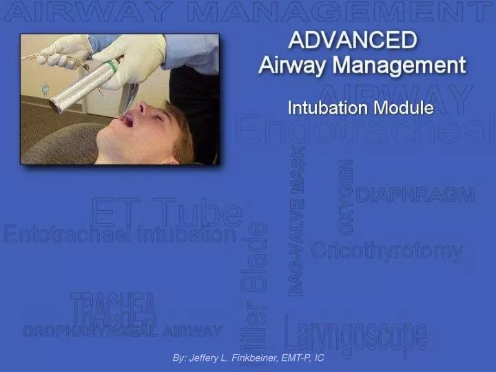
By: Jeffery L. Finkbeiner, EMT-P, IC. Overview. Airway Anatomy Advantages of Intubation Indications Contraindications Complications Equipment Intubation Techniques Nasal Intubation Suctioning. Airway Anatomy. Laryngoscope view of the vocal cords. Advantages of Intubation.

E N D
Overview • Airway Anatomy • Advantages of Intubation • Indications • Contraindications • Complications • Equipment • Intubation Techniques • Nasal Intubation • Suctioning
Airway Anatomy Laryngoscope view of the vocal cords
Advantages of Intubation • A cuffed endotracheal tube protects the airway from aspiration • Access is gained to the tracheobronchial tree for the suctioning of secretions • Ventilations via an entotracheal tube do not cause gastric distention • Maintains a patent’s airway and assists in avoiding further obstruction • Enables delivery of certain medications
Indications • Inadequate oxygenation (decreased arterial PO2) that is not corrected by supplemental oxygen • Inadequate ventilation (increased arterial PCO2) • Need to control and remove pulmonary secretions • Any patient in cardiac arrest • Ant patient in deep coma who cannot protect his airway (without a gag reflex)
Indications, cont... • A patient in immediate danger of upper airway obstruction (i.e. burns of the upper airways) • A patient with a decreased level of consciousness • A patient with severe head and facial injuries with a compromised airway • A patient in respiratory failure or respiratory arrest
Contraindications • A patient with an intact gag reflex • Patients likely to react with laryngospasm (i.e. children with epiglottitis) • Basilar skull fracture (during nasal intubation)
Complications • Trauma to the teeth, vocal cords, soft tissues of the larynx and related structures • Nasotracheal tubes can damage the turbinates, cause severe bleeding, and even perforate the nasopharyngeal membranes • Hypertension and tachycardia can occur from the intense stimulation of intubation. This is potentially life-threatening in the cardiac patient
Complications, cont... • Cardiac arrhythmias related to vagal stimulation or sympathetic nerve stimulation may occur • Damage to the endotracheal tube cuff, resulting in a cuff leak and/or poor seal • Intubation of the esophagus, resulting in gastric distention and regurgitation upon attempting ventilation • Trauma resulting from over ventilating with a BVM without a pressure release valve (pneumothorax)
Complications, cont more... • Over stimulation of the larynx resulting in laryngospasm, causing a complete airway obstruction • Inserting the tube to deeply resulting in right main stem bronchus intubation • Tube obstruction due to foreign material, dried respiratory secretion and/or blood
Equipment Body Substance Isolation (BSI) Face shield/mask, protective glasses and latex examination gloves
Equipment Laryngoscope with relevant size blades, 10cc syringe Miller (straight) Blade MacIntosh (curved) Blade Laryngoscope with blades and 10cc syringe
Equipment Magill Forceps,flexible ET tube stylet Magill Forceps Flexible ET tube stylet Stylet with ET tube
Equipment Relevant size ET tubes, tube holder and/or cloth tape Cuffed and uncuffed ET tubes Various sizes and styles of ET tubes ET tube holder
Equipment BVM with oxygen, suction unit with Yankauer and french ET catheter French ET catheter BVM connected to oxygen Yankauer suction tip
Intubation Techniques • Position yourself at the patient’s head • Inspect the oral cavity for secretions or foreign material. Suction if necessary • Hyperventilate the patient with 100% oxygen for 2 minutes prior to intubation attempt
Intubation Techniques • Place the patient’s head in the sniffing position • Open the patient’s mouth with the fingers of the right hand(easily accomplished by the crossed-fingered technique)
Intubation Techniques • With the laryngoscopeheld in the left hand, insert the blade into the right side of the mouth displacing the tongue to the left • When using a curved blade, advance the tip of the blade into the vallecula (the space between the base of the tongue and the pharyngeal surface of the epiglottis)
Intubation Techniques • When using a straight blade, insert the tip under the epiglottis. The glottic opening is exposed by exerting upward traction on the handle • To allow full visulization of the vocal cords, it may be helpful for an assistant to employ the Selleck’s Maneuver (applying moderate pressure to the cricoid cartilage)
Intubation Techniques • Resist the urge to use a prying motion with the handle. Lift only upward to avoid damaging the patient’s bottom teeth • Advance the ET tube through the right corner of the mouth • Under direct vision, continue advancing the tube through the vocal cords
Intubation Techniques • Holding the tube firmly in place, quickly remove the laryngoscope blade • Observe the depth markings on the the ET tube in relation to the patient’s teeth (19 to 23cm in an adult) • Inflate the cuff with 5 to 10 cc of air via the pre-drawn syringe
Intubation Techniques • Attach the tube to a mechanical ventilation device such as a BVM and begin ventilating and oxygenating the patient • Ensure distal cuff is inflated correctly and observe for any air leaks • Observe end-tidal CO2 monitor and fogging of the tube
Intubation Techniques • During ventilation, confirm proper tube placement • First auscultate the abdomen while visualizing chest expansion • Then auscultate the chest bilaterally ensuring equal breath sounds
Intubation Techniques • Secure the tube in place using a tube holder and cloth tape • If no tube holder is available, the tube may be secured using cloth tape and an oropharyngeal airway • Continue with ventilating the patient
Nasal Intubation Nasal intubation may be indicated for any of the following: • Endotrachael intubation has proven difficult • C-spine motion must be limited (c-spine injury) • If the patient’s jaw is clenched
Nasal Intubation • Hyperventilate the patient with 100% oxygen for 2 minutes prior to intubation attempt • Select a cuffed ET tube 1mm smaller than that used for normal endotracheal intubation • Lubricate the end of the tube with a sterile, water-soluble jelly
Nasal Intubation • Select the nostril that is largest and most direct • With the bevel of the tube toward the septum, advance the tube along the nasal floor
Nasal Intubation • If the nostril is impassible, attempt the other nostril. If unsuccessful, reduce the size of the tube by 0.5mm • The curve of the tube should follow the natural curve of the airway
Nasal Intubation • Gently advance the tube while rotating it medially 15 to 30 degrees • Continue advancing until airflow is heard through the tube • Quickly and gently advance the tube early during the next inspiration (in a non-apnic patient)
Nasal Intubation • Observe for fogging of the tube while advancing. This indicates exhaled breath • If no fogging or breath sounds are are noted, then placement may be in the esophagus. Withdraw and another attempt will be required
Nasal Intubation • Attach the tube to a mechanical ventilation device such as a BVM and begin ventilating and oxygenating the patient • Ensure distal cuff is inflated correctly and observe for any air leaks • Secure the tube with cloth tape
Nasal Intubation • During ventilation observe end-tidal CO2 monitor and confirm proper tube placement • First auscultate the abdomen while visualizing chest expansion • Then auscultate the chest bilaterally ensuring equal breath sounds
Suctioning • It may be necessary to suction secretions from the ET tube • Measure the length of the catheter from the corner of the mouth to the tip of the earlobe • Place your thumb and forefinger on the catheter at the point of maximum depth
Suctioning • Gently insert the catheter into the ET tube (without suction) until you reach the maximum depth determined by your thumb and forefinger • Place your other thumb over the suction hole to begin the vacuum
Suctioning • While holding vacuum, maintain suction while slowly withdrawing the catheter from the tube • Do not suction longer than 15 seconds • Hyperventilate the patient before and after suctioning
This Slide Show is intended for demonstration purposes only.Any other use is unauthorized and is prohibited by law.For information on ordering EMS Continuing Education slidescontact us at:(248) 618-7569orems@twp.waterford.mi.us