Download
1 / 21
220 likes | 661 Views
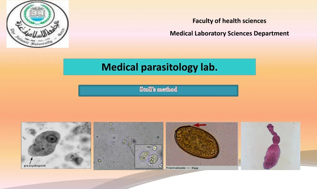
Faculty of health sciences Medical Laboratory Sciences Department. Medical parasitology lab. Stoll’s method. Counting helminthes eggs in feces. The intensity of an intestinal helminthes infection may sometimes be indicated by the concentration of its eggs in feces.

E N D
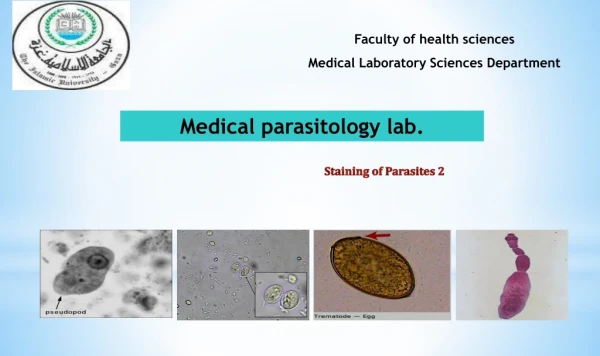
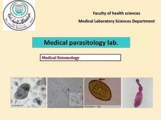
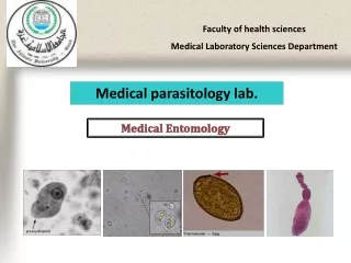
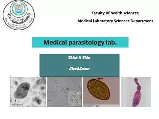
Faculty of health sciences Medical Laboratory Sciences Department Medical parasitology lab. Stoll’s method
Counting helminthes eggs in feces • The intensity of an intestinal helminthes infection may sometimes be indicated by the concentration of its eggs in feces. • Egg count before treatment may help determine whether treatment is needed and counts after treatment assess its success. • Eggs counts can be value in epidemiological surveys. • The approximate number of eggs per gram of feces can be calculated by using formal ether technique. • Egg counts provide a reasonable estimate of the numbers of adult worms present. • When a more accurate count is required the Stoll’s method can be used.
Stool’s technique procedures • Weight 3 grams of feces in a screw cap container. • Add 42 ml of water to give (1/15) dilution of the feces. • If the feces are formed specimen, use sodium hydroxide 0.1mol/l solution instead of water. • Using a rod, break up the feces and mix it with the water. • Cap the container and shake hard to complete in mixing. • Using a graduated plastic bulled pipette, or a Pasteur pipette marked to measure the required volume, pick 150 μl of the suspension and transfer this slide. • Cover the slide with long coverslip if available, or two squares coverslips. • Examine systematically the entire preparation, using 10x objective. Include the count any eggs laying outside the edges of the coverslip because these are contained in 150 μl sample.
8. Multiply the number of eggs counted by 100 to give the number of eggs per gram of feces. If the specimen isn’t formed, the following additional calculation is necessary to give the number of eggs per gram. Fluid specimen -------------------------------- *5 Unformed watery specimen ------------------- *4 Unformed soft specimen ----------------------- *3 Semiformed specimen -------------------------- *2 9. Calculate the number of eggs per day, by multiplying the number of eggs per gram by the total weight of 24hrs fecal specimen.
Calculate the number of burden worm by dividing the number of eggs per gram on number of eggs the parasite laying per day. NOTE: Female Ascaris pass about 200,000 eggs per day, Nectar pass about 9000 eggs per day, and female Trichuris pass about 5ooo eggs per day .
Interpretation of result The Center For Disease Control, Atlanta
Ascaris lumbricoidesgiant round worm Intestinal Nematodes
Ascarislumbricoides Ascaris lumbricoides is the giant roundworm of human, inhabit small intestine and cause Ascariasis . • Infective stage: Embryonated eggs. • There are three diagnostic stages for Ascarislumbricoides in stool: adult, Fertilized eggs, Unfertilized eggs. • DUE TO LARVAL MIGRIATION STAGE, Sputum sample may be collected on persons suffering from infection with Ascarislumbricoid.
Ascaris lumbricoides • Adult (large size ) : • Male: has a coiled posterior end, 2 minute spicules and no copulatory bursa – 15-30 cm in length. • Female: oviparous have straight, pointed tail and no spicules. (lay 200,000 eggs daily) – 20-40 cm in length. Ascaris adult worms are the largest known intestinal nematodes
There are 3 shapes of ova: Embryonated egg Fertilized egg Unfertilized egg embryonated egg
Ascaris lumbricoides Unfertilized Egg Ascarislumbricoides Unfertilized Corticated Egg Ascarislumbricoides Unfertilized deCorticatedEgg
Plant cell resemble helminth eggs particullay Ascaris lumbericoid unfertilized egg. This artifact is typically round to oval in shape and may or may not have a definite cell wall,The interior of the cell looks like a cluster of odd shaped vacuoles. Unfertilized eggs a thin shell protects the inner a morphous mass of protoplasm,The shell layers of the egg provide a very resistant structure which can withstand many chemicals which make them ideal parasites of the intestine.
Ascaris lumbricoides Fertilized Egg Ascarislumbricoides Fertilized corticated egg. Ascaris lumbricoides Fertilized Decorticated Egg
Ascaris lumbricoides Embryonated Egg AscarislumbricoidesEmbryonatedDecorticated Egg AscarislumbricoidesEmbryonatedCorticated Egg
Pollen grain in a concentrated wet mount of stool. This grain looks very similar to the fertile egg of Ascaris lumbricoides, although the spine-like structures on the outer layer should separate the two. fertile egg of Ascaris lumbricoides