Download
1 / 10
100 likes | 283 Views
Prospective Evaluation of Point-of-Care Ultrasonography for the Diagnosis of Pneumonia in Children and Young Adults.

E N D
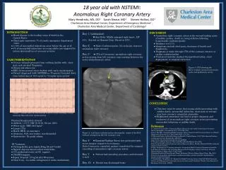
Prospective Evaluation of Point-of-Care Ultrasonography for the Diagnosis of Pneumonia in Children and Young Adults Shah VP, Tunik MG, Tsung JW. Prospective evaluation of point-of-care ultrasonography for the diagnosis of pneumonia in children and young adults. JAMA Pediatr. Published online December 10, 2012. doi:10.1001/2013.jamapediatrics.107.
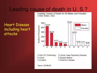
Introduction • Background • Pneumonia is the leading cause of death in children worldwide. • There is poor access to diagnostic imaging worldwide. • Ultrasonography for evaluating pneumonia is feasible and accurate in adults • Study Objective • To determine the accuracy of clinician-performed point-of-care ultrasonography for the diagnosis of pneumonia in children and young adults.
Methods • Study Design • Prospective observational study with a convenience sample of patients. • Setting • Patients presenting to an urban pediatric emergency department who underwent chest radiography for clinical suspicion of pneumonia had lung ultrasonography performed. • Patients • Aged 0-21 years and seen in the emergency department with a clinical suspicion of pneumonia. • Excluded patients who had a previous diagnosis of pneumonia, arrived with a chest radiograph, and had hemodynamic instability.
Methods • Intervention • Enrolled patients had routine examination done with auscultation findings documented and then had lung ultrasonography performed. • Outcomes/Analyses • Clinical follow-up in 2 weeks to determine (1) clinical improvement or deterioration with or without antibiotics and (2) need for unscheduled or repeated emergency department visit. • Limitations • The use of chest radiography as the reference gold standard. • The presence of centrally located pneumonias that did not reach the pleural line and therefore were undetected by ultrasonography.
Results Test Performance Characteristics Using Chest Radiography as a Reference Standard Among 200 Patients
Results Standards for Reporting of Diagnostic Accuracy flowchart.
Results aLung consolidations with sonographic air bronchograms were found measuring between 1.5 and 1.8 cm at the detection limit of chest radiography. bOne of 13 patients had lung consolidation of ≤1 cm without sonographic air bronchograms, indicating consolidation from atelectasis as opposed to pneumonia. Twelve of 13 patients had lung consolidation of ≤1 cm with sonographic air bronchograms, consistent with pneumonia. In addition, Cohen κ was 0.93 (95% CI, 0.87-0.99) between enrolling sonologist interpretation of lung consolidation and expert sonologist review.
Comment • Ultrasonography is: • More specific than sensitive for the detection of pneumonia. • More accurate than World Health Organization–defined tachypnea and auscultation for the diagnosis. • Clinician sonologists were able to accurately and quickly (mean time, 7 minutes) diagnose pneumonia in children. • There was no statistically significant difference in accuracy between sonologists who performed more than 25 lung ultrasonographic examinations and novice sonologists.
Comment • Ultrasonography can detect <1-cm lung consolidations missed by chest radiography. • Point-of-care ultrasonography can be a powerful diagnostic tool to complement the physical examination.
Contact Information • If you have questions, please contact the corresponding author: • James W. Tsung, MD, MPH, Department of Emergency Medicine, Mount Sinai School of Medicine, One Gustave Levy Place, New York, NY 10029 (jtsung@gmail.com). Conflict of Interest Disclosures • None reported.