Download
1 / 59
590 likes | 853 Views
Hemoglobinopathies and Thalassemias. Hemoglobinopathies. Genetically determined abnormalities of the structure or synthesis of hemoglobin molecule. Abnormality associated with globin chain Qualitative defects (structural defect)

E N D
Hemoglobinopathies • Genetically determined abnormalities of the structure or synthesis of hemoglobin molecule. • Abnormality associated with globin chain • Qualitative defects (structural defect) • genetic mutation involving amino acid deletions or substitution • Quantitative defect - thalassemia
Nomenclature of HemoglobinVariants • First discovered was Hemoglobin S (HbS) • Originally, given letter designations beginning with Hemoglobin C (except Hemoglobin F = fetal hemoglobin and Hemoglobin M = hemoglobins that tend to form methemoglobin) • Later given common names according to geographic area in which they were first discovered (e.g. Hb Ft. Worth) • Disease (homozygous) vs. trait (heterozygous)
Pathophysiology • Altered Solubility – when nonpolar amino acid is substituted for a polar amino acid near the surface of the chain (Hb S and C) • Altered Function – polar amino acid substitution for nonpolar residue near the hydrophobic crevice may affect oxygen affinity by stabilizing heme iron in Fe3+ • Altered Stability – substitutions in internal residues may prevent folding into proper tertiary structure
Identification of StructuralHemoglobin Variants • Hemoglobin Electrophoresis • Primary diagnostic tool for differentiating types of qualitative hemoglobinopathies • Separates hemoglobins based on surface charge and movement in an electrical field • Surface charge is affected by the amino acid substitution • Rate of migration depends on support media, pH and ionic strength of buffer, strength of electrical field and time
Cellulose Acetate Electrophoresis • Detection and Preliminary identification of normal and abnormal hemoglobins • Abnormal hemoglobins may require confirmation by citrate agar electrophoresis
Citrate Agar Electrophoresis • Performed at acid pH (6.0) – vs. pH 8.6 for cellulose acetate • Used to differentiate Hg S from D and G • Differentiate C from A2
Thalassemias • Variety of genetic defects in globin chain synthesis – decreased or absent synthesis • Classified according to globin chain that is affected – e.g. β-thalassemia vs. αthalassemia • Heterozygous: minor • Homozygous: major
Pathophysiology • If α chain is affected, excess of β chains produced. If β chain is affected, excess of αchains produced • Imbalance in chain synthesis causes • Decrease in total hemoglobin production • Ineffective erythropoiesis • Chronic hemolysis • Excess α chains are unstable – precipitate within cell – precipitates bind to cell membrane, causing membrane damage
Excess β chains combine to form Hb H (four β chains) • High oxygen affinity – poor oxygen transporter • unstable
Clinical Findings • Anemia/hypoxia • Decreased hemoglobin production • Ineffective erythropoiesis • Presence of high-affinity hemoglobins • Increased extravascular hemolysis • Splenomegaly • Splenic removal of abnormal erythrocytes • Extramedullary hematopoiesis
Gallstones – due to increased intravascular and extravascular hemolysis • Skeletal abnormalities – expansion of bone marrrow • Pathological fractures – thinning of calcified bone • Iron toxicity – multiple transfusions
α-Thalassemia • There are two α genes on each of two chromosome 16 structures (four α genes in the diploid state) • Mutations can affect one or more of the α genes resulting in four levels of severity • When all four genes deleted – no αchains, hydrops fetalis or α-thalassemia major • 3 of the four deleted, hemoglobin H disease
2 of the 4 deleted, α-thalassemia minor • 1 deletion, silent carrier • Primarily affects people of Mediterranean, Asian and African ancestry
Hydrops fetalis • Deletion of all four α genes • No adult hemoglobin can be formed - incompatible with life – infants are stillborn or die within a few hours • Hemoglobin is made using γ, δ and β chains
Hemoglobin H Disease • Usually result when two heterozygous parents (--/αα and the other –α/αα) bear children • Excess of β chains leads to formation of Hb H • At birth, excess of γ chains leads to Hb Bart’s (γ4) • Hb H is unstable – triggering chronic hemolytic anemia • High oxygen affinity
BCB stain in Hb H disease Oxidatively denatured hemoglobin H precipitates
Clinical Findings • Wide variation in degree of anemia • Splenomegaly and hepatomegaly present • Less than ½ of patients exhibit skeletal changes
Laboratory findings • Microcytic/hypochromic anemia (hemoglobin levels 8 to 10 g/dL) • 5-10% reticulocytes • Nucleated red blood cells • 25% Hb Bart’s with levels of Hb A1, Hb A2, and Hb F in neonates • 2-40% Hb H, levels of Hb A2, normal Hb F, remainder Hb A2 in adults
α-Thalassemia minor • Two α genes either on same or opposite chromosomes are missing • Unaffected globin genes are able to compensate for the affected genes • Mild anemia – signficant microcytosis • Normal lifespan
Silent carrier • Affects greater than 25% of African Americans • 3 remaining genes direct synthesis of adequate number of a chains • Totally benign – MCV is borderline (78 –80 fl)
β-thalassemia • Only 2 β globin genes, one on each chromosome 11 • Defect is not deletional • β+ gene mutation causes partial block in βchain synthesis • β0 gene mutation results in complete absence of β chain production • Over 180 mutations resulting in partial to complete absence of β gene expression
β- thalassemia Major –Cooley’s Anemia • Homozygous (β +/ β + or β 0/ β 0) or double heterozygous (β +/ β 0) inheritance • Pathophysiology: dramatic reduction or complete absence of β chain synthesis – • Symptoms begin to manifest at age 6 months • Increase in non β containing hemoglobins • Excess α chains precipitate in cells -hemolysis
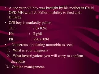
Clinical Symptoms • First observed in infants – irritability, pallor, failure to thrive • Enlarged abdomen • Severe anemia – burdens cardiovascular system- cardiac failure in first decade of life • Growth is retarded; brown pigmentation of skin
Bone changes – facial deformities • Splenomegaly – extramedullary hematopoiesis
Laboratory findings • Hemoglobin as low as 2-3 g/dL • Markedly microcytic/hypochromic • Marked anisocytosis and poikilocytosis • Basophilic stippling and polychromasia • Hemoglobin electrophoresis – 90% Hb F and increased Hb A2
Thalassemia minor syndromes • More common than once thought – • Most common in Mediterranean areas and Asia • Mild compensatory increase in production of chain not affected – e.g. in β -thalassemia minor increase in gamma and delta chains
Thalassemia minor syndromesLaboratory findings • Mild to non-existent anemia • Microcytosis –(hypochromia not striking) • Target cells, basophilic stippling • RDW is normal • Normal iron, ferritin, TIBC
Hemoglobin Electrophoresis • 2-6 % Hgb F (N = < 1% after age 1 year) • 3-7 % Hgb A2 (N = 2-3.5%) • 87-95% Hgb A1 (N=95.5-100%)
Mentzer Index • Calculation that may (or may not) be useful in differentiating thal minor from Fe deficiency • Mentzer Index = MCV/RBC Count • <13 – Thalassemia minor • >13 – Iron Deficiency
Sickle Cell Anemia • Most common symptomatic hemoglobinopathy – highest in Africa • Sickle cell disease in 0.3-1.3% of African Americans; trait in 8-10% of African Americans • HbS in heterozygous state confers advantage against fatal Plasmodium falciparum infections
Pathophysiology of SSA • Mutant hemoglobin (HbS) is produced in which valine (nonpolar) is substituted for glutamine (polar) in 6th position of βchain. (a2b2 6val-glu) • Produces a change in net chg. of molecule; solubility in deoxygenated state is markedly reduced and rigid aggregates of hemoglobin form. • Aggregates polymerize and red cell sickles.
Rate of polymerization depends on • Temperature (temps higher than 37 °C) • pH (acidosis) • Ionic strength (hypertonicity) • Oxygen tension (hypoxia) • Sickled cells return to normal upon reoxygenation – with repeated sickling the membrane undergoes permanent changes and cells become irreversibly sickled
Clinical Findings in SSA • First clinical signs at about 6 month of age • Anemia – moderate to severe anemia as result of extravascular hemolysis • Changes in attempt to compensate for oxygen deficit lead to cardiac overload (cardiac hypertrophy, cardiac enlargement, and congestive heart failure) • Hyperplastic bone marrow (compensation for increased RBC destruction) leads to bone changes
Aplastic crises during or following viral, bacterial, and mycoplasma infections • Vaso-Occlusive Crisis – blocking of microvasculature by rigid sickled cells • Triggered by infection, decreased oxygen, dehydration, slow blood flow, or without any known cause • Pain, low grade fever, organ dysfunction, tissue necrosis
Autosplenectomy– splenic fibrosis and calcification due to infarction • Dactylitis – painful swelling of hand and feet • Bacterial infection – reasons for increased susceptibility not fully understood • Acute splenic sequestration – splenic pooling of sickled RBCs may cause decrease in RBC mass • Acute Chest Syndrome – cough, fever, chest pain, dyspnea, chills, wheezing, pulmonary infiltrates
Laboratory Findings in SSA • Peripheral Blood • Severe anemia (5-9 g/dL) – N/N • Poikilocytosis –sickle cells, target cells • Anisocytosis – Increased RDW • Nucleated RBCs, polychromasia • Leukocytosis (WBC = 12,000-16,000) –absolute neutrophilia with shift to left • Thrombocytosis common – thrombocytopenia during aplastic crises
Electrophoresis on cellulose acetate at pH of 8.4 85 – 100% HbS and <15% HbF • Chemistry tests • Increased bilirubin • Increased LDH • Decreased haptoglobin
Diagnostic Tests for Hgb S • Sickle cell prep Sodium metabisulfite added to blood • Reduces oxygen tension -> sickling • Viewed microscopically • Rare hemoglobin variants may also sickle
Therapy • Preventative – eliminate conditions that precipitate vaso-occulsion • Transfusion during aplastic crises or splenic sequestration • Hydroxyurea to reduce intracellular sickling – reactivated fetal genes and elevated HbF
Sickle Cell Trait • Heterozygous for sickle cell gene • Usually asymptomatic • May have crisis if oxygen tension is sufficiently lowered • Hemoglobin electrophoresis shows 50- 65% HbA1, 35-4% HbS, normal HbF and normal to slightly increased HbA2
Sickle Cell – βThalassemia • Doubly heterozygous • Severity varies from as severe as SSA to asymptomatic • β 0 Thalassemia – no β chain production – more severe • β + Thalassemia – reduced β chain production
Hemoglobin S - β 0 Thalassemia • Many of same findings and crises as in SSA • Hgb from 5-10 g/dL with retic count from 10-20% • Microcytic/hypochromic with marked anisocytosis • Target cells and sickle cells
Hemoglobin S - β + Thalassemia • Hgb will range between 7-10 g/dL to normal. • Few red cell abnormalities • Decrease in MCV or MCH may be only clues to abnormality
Hereditary Persistence of FetalHemoglobin (HPFH) • Group of disorders in which Hgb F production continues throughout life – absence of any significant clinical abnormalities • Heterozygous HPFH – asymptomatic and Hgb F only slightly increased • Homozygous HPFH – microcytosis and mild hypochromasia – no anemia – 100% Hgb F • Important to differentiate thalassemias with high levels of Hgb F from HPHF