Download
1 / 128
1.29k likes | 4.51k Views
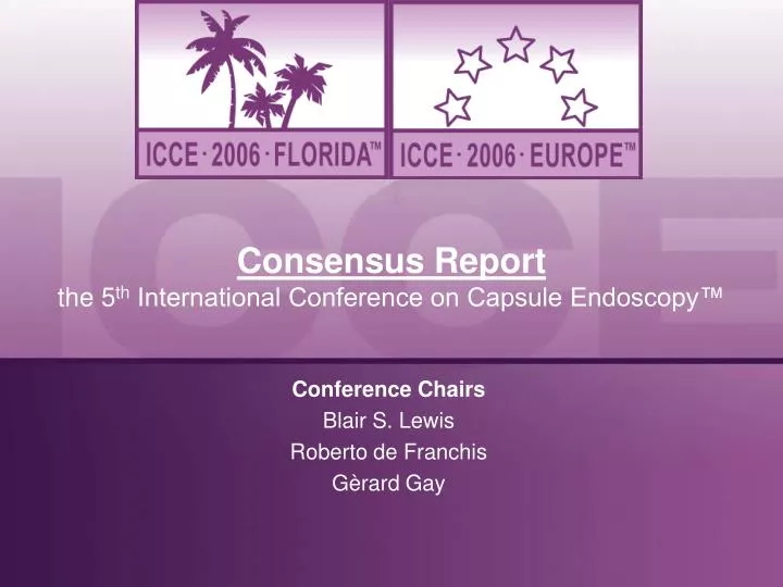
Consensus Report the 5 th International Conference on Capsule Endoscopy™. Conference Chairs Blair S. Lewis Roberto de Franchis Gèrard Gay. ICCE 2006. Two clinical congresses in 2006 Boca Raton, Florida, USA March 6-7, 2006 Paris, France June 9-10, 2006 Combined statistics

E N D
Consensus Reportthe 5th International Conference on Capsule Endoscopy™ Conference Chairs Blair S. Lewis Roberto de Franchis Gèrard Gay
ICCE 2006 • Two clinical congresses in 2006 • Boca Raton, Florida, USA • March 6-7, 2006 • Paris, France • June 9-10, 2006 • Combined statistics • 622 attendees • 40 countries represented • 146 abstracts presented • 89 oral presentations
Consensus Activities • Reviewed last year’s data and updated ICCE 2005 Consensus • Drafted paper for peer-reviewed publication in Endoscopy this fall • Consensus Topics • IBD • Esophagus • Tumors • Bleeding • Celiac • Preps/Prokinetics
Inflammatory Bowel Disease (IBD) June 2006 Panel Co-Chairmen E. Seidman I. Bjarnason Panel Members: J. Leighton, P. Legnani, M. Gassull, J.F. Columbel, V. Manoury, A. Kornbluth
IBD Consensus Capsule Endoscopy (CE) for IBD: • Higher sensitivity for assessing small bowel mucosal lesions compared to other imaging techniques
Meta-analysis of Prospective Comparative Crohn’s Disease Studies: CE vs. Other Modalities 11 studies, n=223 Triester et al Am J Gastroenterol 2006;101:954-964
CE vs. SB Radiography : : Study IY (random) Incremental Yield (random) 95% CI 95% CI Costamagna 2002 0.33 [-0.42, 1.09] Bloom 2003 0.37 [0.08, 0.66] Chong 2003 0.48 [0.22, 0.73] Heigh 2003 0.47 [0.17, 0.77] Buchman 2004 0.00 [-0.27, 0.27] Dubcenco 2004 0.61 [0.42, 0.81] Eliakim 2004 0.54 [0.35, 0.74] Marmo 2004 0.53 [0.26, 0.80] Toth 2004 0.34 [0.17, 0.51] Total (95% CI) 0.42 [0.30, 0.54] Total yield: 66% (CE), 24% (SB radio) Test for heterogeneity: P = 0.03, I² = 52.1% Test for overall effect: P < 0.00001 -1 -0.5 0 0.5 1 Higher yield SB radiography Higher yield CE Triester et al Am J Gastroenterol 2006;101:954-964
CE vs. Ileoscopy Study IY (fixed) IY (fixed) 95% CI 95% CI Bloom 2003 0.05 [-0.26, 0.37] Heigh 2003 0.06 [-0.26, 0.37] Dubcenco 2004 0.32 [0.09, 0.55] Toth 2004 0.11 [-0.09, 0.30] Total (95% CI) 0.15 [0.02, 0.27] Total yield: 61% (CE), 46% (Ileoscopy) Test for heterogeneity: P = 0.38, I² = 2.1% Test for overall effect: P = 0.02 -1 -0.5 0 0.5 1 Higher yield Ileoscopy Higher yield CE Triester et al Am J Gastroenterol 2006;101:954-964
CE vs. CT Enterography (CTE) Study IY (fixed) IY (fixed) 95% CI 95% CI Heigh 2003 0.18 [-0.14, 0.50] Voderholzer 2003 0.00 [-0.42, 0.42] Eliakim 2004 0.57 [0.38, 0.76] Total (95% CI) 0.38 [0.23, 0.54] Total yield: 75% (CE), 37% (CTE) Test for heterogeneity: P = 0.01, I² = 76.2% Test for overall effect: P < 0.00001 -1 -0.5 0 0.5 1 Higher yield CTE Higher yield CE Triester et al. Am J Gastroenterol 2006;101:954-964
Summary of Incremental Yield (IY) of CE Over Other Modalities Triester et al. Am J Gastroenterol 2006;101:954-964
CE vs. Barium Radiography Suspected CD subgroup Study IY (random) [95% CI] IY (random) [95% CI] 0.00 [-0.85, 0.85] Costamagna 2002 0.38 [-0.04, 0.79] Dubcenco 2004 0.54 [0.35, 0.74] Eliakim 2004 0.17 [-0.02, 0.37] Toth 2004 Chong 2005 0.00 [-0.11, 0.11] Hara 2005 0.25 [-0.16, 0.66] Total (95% CI) 0.24 [-0.03, 0.51] Total yield (fixed): 43% (CE), 13% (barium radiography) Test for heterogeneity: P < 0.001, I² = 85.6% Test for overall effect: P = 0.09 0 1 0.5 -1 -0.5 Yield higher in barium radiography Yield higher in capsule endoscopy Established CD subgroup Study IY (random) [95% CI] IY (random) [95% CI] 0.50 [-0.21, 1.21] Costamagna 2002 0.03 [-0.20, 0.27] Buchman 2004 0.70 [0.49, 0.90] Dubcenco 2004 0.45 [0.23, 0.67] Marmo 2004 0.61 [0.35, 0.87] Toth 2004 Chong 2005 0.62 [0.38, 0.86] Hara 2005 0.67 [0.34, 0.99] Total (95% CI) 0.51 [0.31, 0.70] Total yield (fixed): 78% (CE), 32% (barium radiography) Test for heterogeneity: P = 0.001, I² = 72.9% Test for overall effect: P < 0.001 -1 -0.5 0 0.5 1 Yield higher in barium radiography Yield higher in capsule endoscopy
CE vs. CT Enterography (n=58 pts) CE detects more proximal disease + exams Voderholzer et al. Gut 2005;54:369-373 Hara et al. Radiology 2006;238(1):128-134
MR Enteroclysis (n=18 pts) + exams Golder et al. Int’l J of Colorectal Disease 2006;21(2):97-104
IBD Consensus Capsule endoscopy (CE) vs. other imaging: • Limitations • The available data are more evidence based for known, non-stricturing CD than for suspected CD. • No “gold standard” available for CD. • CE is superior to CT enterography & MRI; particularly for proximal - mid small bowel CD. • CE demonstrates mucosal lesions missed by other imaging. • No single test is available for diagnosing CD.
IBD Consensus CE may be useful in the study of indeterminate colitis: • 22 pts with colonic IBD underwent CE. • 9 (40%) with “colitis” were found to have small bowel lesions. • 27 pts with IC underwent CE. • 8 (29%) had small bowel lesions. • 10 pts with IC underwent CE. • 4 (40%) had small bowel lesions. Mow WS, et al. CGH 2004;2:31-40 Mascarenhas-Saraiva M, et al. ICCE 2005 AB 115 Hume G, et al. ICCE 2004 AB 1054
IBD Consensus • 31 patients with IC and known serology • CE and serology equally sensitive (61%). • CE was more sensitive than ASCA or OMP-C in diagnosing small bowel CD. • Conclusion:CE was superior to CD-like markers in identifying small bowel disease in IC patients. Lo SK, et al., Gastrointest Endosc 2003;57(5):AB 1889
IBD Consensus Role of CE in assessing for early post- operative recurrence • 32 post-op ileocecal resection • CE & ileo-colonoscopy < 6 months • Recurrence: 21/32 – sensitivity • Ileo-colonoscopy 90% vs. 62% for CE • CE identified more proximal disease in 2/3 of cases. • CE may be useful as a first line evaluation of post-operative recurrence due to its good tolerability. Bourreille et al Gut 2006;55:978-983
IBD Consensus Role of CE in assessing for early post-operative recurrence • 14 patients post-op ileocecal resection x 1 yr • CE & small bowel US compared in 13 (1 stricture) • Recurrence: 12/13 by colonoscopy • US: 13/13 ( 1 false +) • CE: 12/13 (all true +) • CE represents an alternative minimally-invasive technique for assessing CD recurrence in patients under follow-up of ileo-colonic resection. Biancone et al; Gastroenterology 2006;130(4):Supp S2: AB S1336
IBD Consensus Capsule endoscopy (CE) for suspected IBD: • Useful and safe in patients with suspected Crohn’s disease and negative endoscopic & small bowel imaging • Evidence: based mainly on retrospective studies; more prospective data needed. • Positive CE findings not well defined (lack of validated scoring index). • Has potential to affect patient management. • Scoring index may provide diagnostic threshold.
Capsule Endoscopy: Are All Ulcers Crohn’s? A B C Which image is an ulcer from Crohn’s disease? The answer is all three. However, patient history will define if another cause, such as NSAID damage or radiation enteropathy caused the ulceration.
IBD Consensus • Standardized CE scoring index of disease severity to differentiate normal from small bowel inflammatory disorders in development. • Correlation of CE index with clinical disease activity scores needed. • CE scoring index may not distinguish between various causes of inflammation (NSAIDs, radiation enteropathy).
Scoring Index Parameters Villous Appearance Ulceration Stenosis Scale Normal, edematous Number - single, few, multiple Distribution - localized, patchy, diffuse Longitudinal extent - short, long, whole segment Ulcer size - based on amount of bowel wall circumference involved Stenosis - ulcerated or not, traversed or not
Example of Score Template Global Disease Assessment: Normal, Mild, Moderate/Severe
Suspected Crohn’s Disease Patients with characteristic GI symptoms of CD (at least 1 from “A”), and with at least one of the criteria under “B”, “C” or “D”: Characteristic GI Symptoms (anti-tTG negative) Chronic abdominal pain Chronic diarrhea Significant weight loss Growth failure Extra-intestinal Symptoms Unexplained recurrent fever Arthritis/arthralgias Pyoderma/erythema nodosum Aphthous stomatitis Perianal disease PSC/recurrent cholangitis Inflammatory Markers Iron deficiency anemia Thrombocytosis or leukocytosis Elevated ESR or CRP Hypoalbuminemia Positive IBD serology Fecal markers: lactoferrin, alpha-1 antitrypsin, calprotectin; heme +; leucocyte + Abnormal, Non-diagnostic Imaging
Suspected SB CD Positive ileocolonoscopy Negative ileocolonoscopy or unsuccessful No obstruction Possible or known obstruction Patency capsule either/or No obstruction Obstruction Capsule endoscopy CTE/MRE (SBFT) Presence of SBCD Treat accordingly Figure 1. Algorithm for the approach to suspected small bowel Crohn’s Disease (CD). The absence of any mucosal lesions demonstrated by a complete assessment of the small bowel by capsule endoscopy excludes active CD of the small bowel. Patients with symptoms suggestive of obstruction, or known to have a stenosis should either undergo a patency capsule exam or evaluation by CTE or MRE prior to capsule endoscopy. Abbreviations: SB CD=small bowel Crohn’s Disease, CTE=CT enterography, MRE=MR enterography, SBFT=small bowel follow through.
Capsule Retention in Crohn’s Disease • In patients with Established CD, the risk is 5%, despite absence of strictures on SBFT. • In cases with Suspected CD: • The risk is low with negative SBFT. • If no SBFT, in the absence of obstructive symptoms, risk is yet unknown.
Conclusions • CE has a higher sensitivity for assessing small bowel mucosal lesions compared to other imaging techniques. • CE is helpful diagnosing suspected Crohn’s in the pediatric population. • CE is superior to CT enterography & MRI; particularly for proximal - mid small bowel CD. • CE may be useful as a first line evaluation of postoperative recurrence of CD. • CE can detect small bowel lesions in a significant number of patients with indeterminate colitis and may alter disease management. • CE is useful and safe in patients with suspected Crohn’s disease and negative endoscopic & small bowel imaging.
Esophagus June 2006 Panel Co-Chairmen R. Eliakim G. Eisen Panel Members: J.P. Galmiche, T. Roesch, F. Schnoll-Sussman, J. Herrerias, V.K. Sharma, E. Coron
Consensus Statement - Esophageal Capsule Endoscopy (ECE) Esophageal Varices Barrett’s Esophagus • A new approach to esophageal diagnostics • Simple and easy • Patient-friendly • Screening tool for esophageal diseases • Encouraging initial clinical data
Consensus Statement – Varices • Esophageal varices (EV) are a serious consequence of portal hypertension (PHT). • In patients with cirrhosis, the incidence of EV increases 5% per year and the rate of progression from small to large varices is 5-10%. • Increasing size of varices is associated with increased wall tension leading to rupture and bleeding. • AASLD/UK guidelines recommend endoscopic screening of patients with cirrhosis for varices and treatment of patients with medium/large varices to prevent bleeding. Eisen G, De Franchis R, Eliakim R, Zaman A, Schwartz J, Faigel D, Rondonotti E, Villa F, Weizman E, Yassin K. Preliminary results of International Multicenter Trial. 32 patients reported. ICCE 2006 AB 20154
Consensus Statement – Varices (continued) • Recommended endoscopic screening intervals are 1-3 years, depending on presence/absence of varices and whether patient has compensated/decompensated liver disease. • Endoscopic surveillance is performed in patients after obliteration of varices. • This patient population could benefit from a non-invasive diagnostic test that does not require sedation. • These recommendations/practices represent a potentially large endoscopic burden. Eisen G, De Franchis R, Eliakim R, Zaman A, Schwartz J, Faigel D, Rondonotti E, Villa F, Weizman E, Yassin K. Preliminary results of International Multicenter Trial. 32 patients reported. ICCE 2006 AB 20154
EV Screening Pilot Trial • Initial pilot trial – EV screening with ESO • Prospective blinded, 3 center study • 32 patients – enriched population with surveillance • No complications, no retention • Japanese endoscopic grading system • F0 = none • F1 = small • F2 = medium • F3 = large • Modified classification for current trial • None/small/medium-large • Medium-Large > 25% circumference Eisen G, Eliakim R, Zaman A, Schwartz J,Faigel D, Rondonotti E, Villa F, Weizman E, Yassin K, de Franchis R. Endoscopy 2006:38:1-5
Comparison of PillCam ESO and EGD: Esophageal Varices 1.Lapalus MG. Endoscopy 2006;38:36-4 2. Eisen GM, de Franchis R. Interim Analysis of the Evaluation of PillCam ESO in the Detection of Esophageal Varices AB 20154 3.Eisen G, de Franchis R, Eliakim R, Zaman A, Schwartz J, Faigel D, Rondonotti E, Villa F, Weizman E, Yassin K, Endoscopy 2006;38(1):1-5
Epidemiologyin Barrett’s Esophagus 7% of US Population have daily GERD Symptoms 10% of Chronic GERD Patients have Barrett’s esophagus Risk of esophageal cancer in Barrett’s esophagus 30-60 times > general population up to 2% of patients with BE Locke III et al. Gastro 1997: 112:1448-1456. Falk GW. Gastro Endosc 1999; 49(3):S29-34.
Screening for Barrett’s Esophagus • Adenocarcinoma is a lethal disease. • GERD is a firmly established risk factor for this cancer. • Barrett’s esophagus, a premalignant precursor, is firmly associated with GERD symptoms, and is clearly associated with an increased risk of cancer (RR 30-60 X general population).
Multi-center Study Overview • Primary aims • Accuracy of ECE compared with EGD for the diagnosis of esophageal pathology in patients with chronic GERD symptoms • Specificity, sensitivity, PPV, NPV • Safety and adverse events of ECE • Secondary aims • Assess capability of ECE to identify presence of Barrett’s esophagus in patients undergoing surveillance endoscopy • Assess patient satisfaction with both procedures • Multi-site: Prospective 7-center international study • Israel (3), USA (3), Germany (1) • Inclusion criteria • Aged 18 years or older • Confirmation of 1 of the following: • Histologic confirmation of Barrett's esophagus undergoing surveillance endoscopy • Chronic GERD symptoms undergoing upper endoscopy for the evaluation of GERD Eliakim R et al. J Clin Gastroenterol 2005;39:572-578
Patient Enrollment 109 patients enrolled 1 unable to swallow capsule 2 technical difficulties 106 included in per-protocol statistical analysis 93 (88%) endoscoped for GERD symptoms 13 (12%) for surveillance of Barrett’s esophagus Eliakim R et al. J Clin Gastroenterol 2005;39:572-578
Methods • ECE swallowed using standardized ingestion protocol. • Blinded investigator reviewed ECE videos. • Upper endoscopy performed on the same day following ECE. • Adjudication committee arbitrated if discrepancy between procedures was noted. • Barrett’s cases were not biopsied for confirmation. Eliakim R et al. J Clin Gastroenterol 2005;39:572-578
Multi-center Study Results:Esophagitis Adjudicated results Eliakim R, Sharma VK et al. In press. J Clin Gastro
Multi-center Results:Barrett’s Esophagus Adjudicated results Eliakim R, Sharma VK et al. In press. J Clin Gastro
ECE Clinical Trial Data:Barrett’s Esophagus 1.Lin et al. Blinded Comparison of Esophageal Capsule Endoscopy vs. Conventional Endoscopy for Diagnosis of Barrett’s Esophagus in Patients with Chronic Gastroesophageal Reflux GIE ( in Press) 2.Sharma et al Gastroenterology 2006;130(4) April AB S1812
Conclusions • ECE does offer a minimally invasive method to screen for esophageal varices and portal hypertensive gastropathy. • ECE does have a role in the evaluation of patients with esophageal disease that would otherwise avoid traditional testing methods. • Large scale studies are needed to confirm outcomes.
GI Bleeding June 2006 Panel Co-Chairmen M. Pennazio I. Gralnek Panel Members: M. Delvaux, N. Reddy S. Bar Meir, I. Demedts, M. Keuchel
Panel Participants(Boca Raton/Paris) Martin Keuchel Ingrid Demedts Simon Bar-Meir Nageshwar Reddy Michael Delvaux Scott Ketover Morry Moskovitz Shenan Abey Colm O’Morain
Value of CE for Obscure GI Bleeding • CE is a valuable diagnostic modality in evaluating obscure GI bleeding. • Key advantages of CE include: ability to image entire small bowel; ability to review and share images; patient preference; safety profile; ability to conduct in variety of settings; clarity of image comparable to other endoscopy. • 2 meta-analyses support role of CE in OGIB*. *Triester et al. Am J Gastro 2005;100:2407-2418 *Marmo et al. APT 2005;22:595-604
Value of CE for Obscure GI Bleeding Marmo et al. APT 2005;22:595-604