Download
1 / 30
350 likes | 793 Views
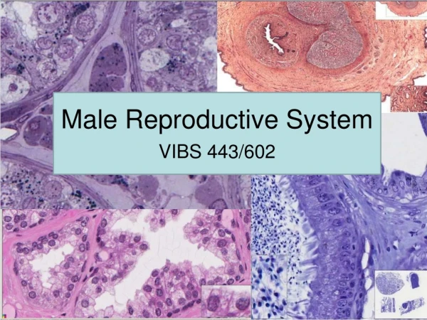
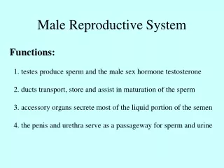
MALE REPRODUCTIVE SYSTEM. Dr Iram Tassaduq. COMPONENTS. The male reproductive system consists of the testes, genital ducts and accessory sex glands . The accessory sex glands include the seminal vesicles, prostate, & bulbouretheral glands. . FUNCTIONS.

E N D
MALE REPRODUCTIVE SYSTEM Dr Iram Tassaduq
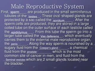
COMPONENTS • The male reproductive system consists of the testes, genital ducts and accessory sex glands . • The accessory sex glands include the seminal vesicles, prostate, & bulbouretheral glands.
FUNCTIONS • The testes have two functions: they produce the male gametes or spermatozoa, and they produce male sexual hormone, testosterone, which stimulates the accessory male sexual organs and causes the development of the masculine sex characteristics.
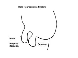
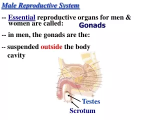
TESTIS • The adult testes are paired ovoid organs that lie within the scrotum, located outside the body cavity. • Testes are suspended by the spermatic cords and tethered to the scrotum by scrotal ligaments, the remnants of the gubernaculum.
STRUCTURE OF TESTIS • The testes have an unusually thick connective tissue capsule known as tunica albuginea. • The inner part of this capsule, the tunica vasculosa, is a loose connective tissue layer that contains blood vessels.
Along the posterior surface of testis, the tunica albuginea thickens and project inwards as the medistinum testis.
LOBULES OF TESTES • From the mediastinum, delicate fibrous septa radiate towards the tunica albuginea and divide the parenchyma of the testis into about 300 lobules, which communicate peripherally. Each lobule contains 1-4 convoluted seminiferous tubules
SEMINIFEROUS TUBULE • Each seminiferous tubule continues near the mediastinum into a straight tubule, a tubulus rectus. The straight tubules continue into the rete testis.
CELLS OF SEMINIFEROUS TUBULE • The seminiferous epithelium is an unusual and complex striated epithelium composed of two basic cell populations: • Sertoli cells (supporting or Sustentacular cells) • Spermatogenic cells
SEMINIFEROUS EPITHELIUM 1. Spermatogonia 2. Primary spermatocytes 3. Spermatids 4. Developing spermatozoa 5. Sertoli cell nucleus 6. Myoepithelial cell of tunica propria 7. Interstitial cell of Leydig 7
SEMINIFEROUS EPITHELIUM • The most immature spermatogenic cells, called spermatogonia, rest on the basal lamina. • The most mature cells, called spermatids, are attached to the apical portion of the Sertoli cell, where they border the lumen of the tubule.
SPERMATOGENIC CELLS • These cells regularly replicate and differentiate into mature sperm. • These cells are derived from primordial germ cells originating in the yolk sac. • Spermatogenic cells are organized in poorly defined layers of progressive development between adjacent Sertoli cells.
SERTOLI CELLS • are far less numerous than the spermatogenic cells and are evenly distributed between them. Their shape is highly irregular
SERTOLI CELLS • Also known as supporting, or sustentacular cells. • These cells do not replicate after puberty. • Sertoli cells are columnar cells with extensive apical and lateral processes.
FUNCTIONS • Support, protection and nutritional regulation of spermatozoa • Phagocytosis • Secretion • Production of AMH • Formation of blood testes barrier
LEYDIG CELLS Leydig cells (interstitial cells) are large polygonal eosinophilic cells that contain lipid droplets. Lipofuscin pigment is also frequently present in these cells as well as distinctive, rod shaped cytoplasmic crystals, the crystals of Reinke.
TUNICA (LAMINA) PROPRIA • Also called peritubular tissue. • This is a multilayered connective tissue that lacks typical fibroblasts. • In man, it consists of 3 to 5 layers of myoid cells (peritubular contractile cells)and collagen fibrils, external to the basal lamina of the seminiferous epithelium.
DIAGRAM OF THE TUBULAR ARCHITECTURE Adluminal compartment Spermatids The blood testis barrier Primary spermatocyte Intercellular bridge Junctional complex Basal compartment Spermatogonium Basement membrane Interstitial compartment Leydig cell Blood vessel
DEVELOPMENT OF TESTIS • The testes develop on the posterior wall of the abdomen and later descend into the scrotum. • Testis are derived from 3 sources: • Intermediate mesoderm • Mesodermal epithelium • Primodial germ cells