Download
1 / 20
200 likes | 316 Views
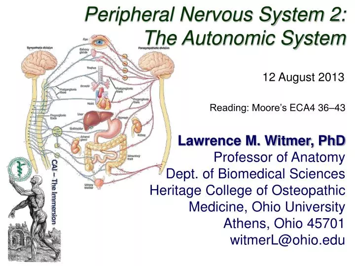
Peripheral Nervous System 2: The Autonomic System. 12 August 2013. Reading: Moore’s ECA4 36–43. Lawrence M. Witmer, PhD Professor of Anatomy Dept. of Biomedical Sciences Heritage College of Osteopathic Medicine, Ohio University Athens, Ohio 45701 witmerL@ohio.edu.

E N D
Peripheral Nervous System 2: The Autonomic System 12 August 2013 Reading: Moore’s ECA4 36–43 Lawrence M. Witmer, PhD Professor of Anatomy Dept. of Biomedical Sciences Heritage College of Osteopathic Medicine, Ohio University Athens, Ohio 45701 witmerL@ohio.edu
Somatic vs. Visceral Langman’s Embryo 9 2004
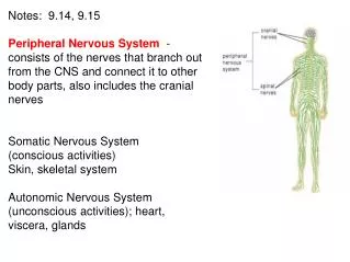
Sensory/Motor + Somatic/Visceral Somatic Nervous System Autonomic Nervous System (July 29) (today)
Overview of the Autonomic Nervous System Similarities between Sympathetic & Parasympathetic • Both are efferent (motor) systems: “visceromotor” • Both involve regulation of the “internal” environment generally outside of our conscious control: “autonomous” • Both involve 2 neurons that synapse in a peripheral ganglion • Innervate glands, smooth muscle, cardiac muscle glands ganglion CNS smooth muscle cardiac muscle preganglionic neuron postganglionic neuron
Overview of the Autonomic Nervous System Differences between Sympathetic & Parasympathetic Location of Preganglionic Cell Bodies Sympathetic Parasympathetic Thoracolumbar T1 – L2/L3 levels of the spinal cord Craniosacral Brain: CN III, VII, IX, X Spinal cord: S2 – S4
Overview of the Autonomic Nervous System Differences between Sympathetic & Parasympathetic Relative Lengths of Neurons Sympathetic target ganglion CNS short preganglionic neuron long postganglionic neuron Parasympathetic target ganglion CNS long preganglionic neuron short postganglionic neuron
Overview of the Autonomic Nervous System Differences between Sympathetic & Parasympathetic Neurotransmitters Sympathetic NE (ACh at sweat glands), + / -, α & ß receptors ACh, + • All preganglionics release acetylcholine (ACh) & are excitatory (+) • Symp. postgangl. — norepinephrine (NE) & are excitatory (+) or inhibitory (-) • Parasymp. postgangl. — ACh & are excitatory (+) or inhibitory (-) • Excitation or inhibition is a receptor-dependent & receptor-mediated response Parasympathetic ACh, + Potential for pharmacologic modulation of autonomic responses ACh, + / - muscarinic receptors
Overview of the Autonomic Nervous System Differences between Sympathetic & Parasympathetic Target Tissues Sympathetic Parasympathetic • Organs of head, neck, trunk, & external genitalia • Organs of head, neck, trunk, & external genitalia • Adrenal medulla • Sweat glands in skin • Arrector muscles of hair • ALL vascular smooth muscle » Sympathetic system is distributed to essentially all tissues (because of vascular smooth muscle) » Parasympathetic system never reaches limbs or body wall (except for external genitalia)
Overview of ANS Functional Differences Sympathetic • “Fight or flight” • Catabolic (expend energy) Parasympathetic • “Feed & breed”, “rest & digest” • Homeostasis » Dual innervation of many organs — having a brake and an accelerator provides more control
Structure of spinal nerves: Somatic pathways dorsal ramus dorsal root ganglion dorsal root spinal nerve somatic sensory nerve (GSA) dorsal horn CNS inter- neuron somatic motor nerve (GSE) ventral horn ventral ramus gray ramus communicans ventral root white ramus communicans Mixed Spinal Nerve sympathetic ganglion
Structure of spinal nerves: Sympathetic pathways dorsal ramus intermediolateral gray column spinal nerve ventral ramus gray ramus communicans white ramus communicans sympathetic ganglion
Sympathetic System: Preganglionic Cell Bodies • Preganglionic cell bodies in intermediolateral gray • T1 – L2/L3 • Somatotopic organization somatic tissues (body wall, limbs) visceral tissues (organs) intermediolateral gray columns T1 – L2/L3 lateral horn Clinical Relevance » dysfunction due to cord injury » spinal nerve impingement & OMM » referred pain Moore’s COA6 2010
Sympathetic System: Postganglionic Cell Bodies 1. Paravertebral ganglia • Located along sides of vertebrae • United by preganglionics into Sympathetic Trunk • Preganglionic neurons are thoracolumbar (T1–L2/L3) but postganglionic neurons are cervical to coccyx • Some preganglionics ascend or descend in trunk Paravertebral ganglia sympathetic trunk (chain) synapse at same level Prevertebral ganglia • celiac ganglion • sup. mesent. g. • inf. mesent. g. ascend to synapse at higher level descend to synapse at lower level aorta Moore’s COA6 2010
Sympathetic System: Postganglionic Cell Bodies 2. Prevertebral (preaortic) ganglia • Located anterior to abdominal aorta, in plexuses surrounding its major branches • Preganglionics reach prevertebral ganglia via abdominopelvic splanchnic nerves Paravertebral ganglia sympathetic trunk (chain) Prevertebral ganglia • celiac ganglion • sup. mesent. g. • inf. mesent. g. abdominopelvic splanchnic nerve aorta Moore’s COA6 2010
Sympathetic System: Summary visceral tissues (organs) Cardiopulmonary Splanchnics: postganglionic fibers to thoracic viscera somatic tissues (body wall, limbs) T1 postganglionics via 31 spinal nerves to somatic tissues of neck, body wall, and limbs Abdominopelvic Splanchnics: preganglionic fibers to prevertebral ganglia, postganglionic fibers to abdominopelvic viscera sympathetic trunk L2 prevertebral ganglia Moore’s COA6 2010
Parasympathetic Pathways Cranial outflow • CN III, VII, IX, X • Four ganglia in head • Vagus nerve (CN X) is major preganglionic parasymp. supply to thorax & abdomen • Synapse in ganglia within wall of the target organs (e.g., enteric plexus of GI tract) Sacral outflow • S2–S4 via pelvic splanchnics • Hindgut, pelvic viscera, and external genitalia Clinical Relevance » Surgery for colorectal cancer puts pelvic splanchnics at risk » Damage causes bladder & sexual dysfunction Moore’s COA6 2010
Visceral Afferents and Referred Pain dorsal root ganglion Visceral sensory nerves [GVA] • run with sympathetic & parasympathetic nerves • cell bodies in dorsal root ganglion • nerve ending in viscera Somatic sensation: • conscious, sharp, well-localized • touch, pain, temperature, pressure, proprioception Visceral sensation: • often unconscious; if conscious: dull, poorly-localized • distension, blood gas, blood pressure, cramping, irritants
Visceral Afferents and Referred Pain Referred Pain: • Pain originating in a visceral structure perceived as being from an area of skin innervated by the same segmental level as the visceral afferent • Results from convergence of somatic & visceral afferents on the same segmental level of the spinal cord • “Cross-talk” in the dorsal horn convergence & “cross-talk” somatic afferent www.merck.com visceral afferent Kandel et al. 2000
Visceral Afferents and Referred Pain Maps of Referred Pain Grant’s Atlas 122009
References Agur, A. M. R. and A. F. Dalley. 2009. Grant’s Atlas of Anatomy, 12th Edition. Lippincott, Williams & Wilkins, New York. Kandel, E. R., J. H. Schwartz, and T. M. Jessell. 2000. Principles of Neural Science, 4th Edition. McGraw-Hill, New York. Moore, K. L., A. F. Dalley and A. M. E. Agur. 2010. Clinically Oriented Anatomy, 6th Edition. Lippincott, Williams & Wilkins, New York. Sadler, T. W. 2004. Langman’s Medical Embryology, 9th Edition. Lippincott, Williams & Wilkins, New York.