Download

1 / 13
150 likes | 275 Views
CLASSIFICATION OF ANEMIA. Prof. Dr. S. Sami Kartı. CLASSIFICATION OF ANEMIA Morphologic Approach.

E N D
CLASSIFICATION OF ANEMIA Prof. Dr. S. Sami Kartı
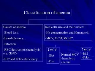
CLASSIFICATION OF ANEMIAMorphologic Approach • The morphologic approach to anemia begins with review of the CBC, particularly the mean corpuscular volume (MCV), and the peripheral blood smear. The initial distinction is based on the red cell size: anemias are classified as microcytic, normocytic, or macrocytic • The presence of abnormally shaped erythrocytes (poikilocytes) may suggest a specific disease or cause • A problem with the morphologic approach is that the morphologic changes in early anemia may be subtle and easy to miss • A second problem is that one morphologic abnormality may have several possible causes
Classification of Anemias (Morphologic) • Microcytic anemias • Iron deficiency • Thalassemia • Sideroblastic anemia • Anemia of chronic diseases (severe cases) • Normocytic anemias • Anemia of chronic diseases (most cases) • Iron deficiency (early) • Anemia of renal disease • Combined nutritional deficiencies (iron + folate or cobalamine) • Marrow failure • Hypothyroidism • Macrocytic anemias • Megaloblastic anemia (folate or cobalamine deficiency) • Hemolytic anemia (reticulocytosis) • Liver disease • Hypothyroidism • Myelodysplasia
CLASSIFICATION OF ANEMIAPathophysiologic (Functional or Kinetic) Approach • The pathophysiologic approach is based primarily on the reticulocyte count • Anemias are classified into three broad categories • Hypoproliferative anemias • Maturation defects • Hemorrhagic/hemolytic (hyperproliferative) anemias
Hypoproliferative anemias • The marrow fails to appropriately respond to the anemia, but the cells that are produced are usually normal • The reticulocyte count or reticulocyte production index is low • Erythrocyte morphology is unremarkable • Anemias in this group are • Iron deficiency anemia • Anemia of chronic disease • Anemias caused by decreased erythropoietin production (chronic renal disease, endocrine disorders) • Anemias caused by marrow damage (stem cell damage, structural damage, autoimmune)
Marrow Damage Anemias • Stem cell damage • Chemotherapy • Drugs: antibiotics, antidepressants • Chemicals: solvents, heavy metals • Infections: bacterial or viral • Aplastic anemia • Structural damage • Radiation • Metastatic malignancies • Myelofibrosis • Granulomatous diseases: tuberculosis, fungal diseases • Storage diseases: Gaucher’s disease • Autoimmune or unknown • Infections: hepatitis, Ebstein Barr virus, HIV • Rheumatic disorders: Systemic Lupus Erythematosis (SLE) • Aplastic anemia/pure red cell aplasia • Graft versus host disease • Congenital: Blackfan-Diamond
Maturation defect anemias • The bone marrow is attempting to respond to the anemia, but the cells produced are unable to enter the circulation and most die within the bone marrow (ineffective erythropoiesis) • The reticulocyte count is low, and erythrocyte morphology is abnormal • The maturation defect anemias are subclassified into • cytoplasmic maturation defects, which are generally associated with microcytic erythrocytes (severe iron deficiency, thalassemia, sideroblastic anemia) • nuclear maturation defects, which are associated with macrocytic erythrocytes (megaloblastic anemias such as Vit. B12 deficiency or folic acid deficiency, myelodysplasia which is an intrinsic marrow disease)
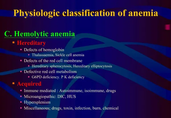
Hemorrhagic/hemolytic (hyperproliferative) anemias • There is increased destruction or loss of erythrocytes • The bone marrow is attempting to respond to the anemia and is producing mature erythrocytes but is unable to fully compensate for the increased red cell loss • The reticulocyte production index is high (>3) and the MCV is frequently high since reticulocytes are larger than normal mature erythrocytes • Anemias in this group are • Acute blood loss • Acute hemolysis (intravascular, extravascular) • Chronic hemolysis (environmental disorders, membrane defects, metabolic defects, hemoglobinopathies, paroxysmal nocturnal hemoglobinuria-PNH)
Evaluation of a Microcytic Anemia (MCV < 80 fL) (I) • The key initial steps in the evaluation of a microcytic anemia are iron indices and examination of a blood smear • The most common cause of microcytic anemia is iron deficiency. If the iron indices confirm the presence of an iron deficiency, the next step is to discover the cause (blood loss, insufficient dietary iron) and begin replacement therapy • If the iron studies do not suggest iron deficiency, the next step is to order a hemoglobin electrophoresis to diagnose β-thalassemia (increased hemoglobin A2) or a hemoglobinopathy • α-Thalassemia is usually diagnosed largely by exclusion (a microcytic anemia in the absence of iron deficiency and decreased hemoglobin A2 is most likely α-thalassemia)
Evaluation of a Microcytic Anemia (MCV < 80 fL) (II) • Consider the ethnic origin and family history of the patient. A blood smear could be done to check for target cells and basophilic stippling. Complete blood counts and blood smears from relatives might be helpful in this circumstance • Consider the possibility of a chronic inflammatory process that might be causing anemia of chronic disease • If none of these appear to be responsible for the anemia, a bone marrow examination with an iron stain to look for ringed sideroblasts might be required
Evaluation of a Macrocytic Anemia (MCV > 100 fL) (I) • The most important initial step in the evaluation of an anemia with an increased MCV is to differentiate megaloblastic anemia from macrocytic, non-megaloblastic anemia • Examine a blood smear for hypersegmented neutrophils and oval macrocytes, which would suggest a megaloblastic anemia • The MCV can also prove helpful; the MCV in megaloblastic anemia is often ≥120 fL, whereas in non-megaloblastic anemias, it is usually ≤115 fL • The first laboratory studies should be serum cobalamin, serum folate, and red cell folate levels. If one of these is abnormal, the cause must be determined and therapy started
Evaluation of a Macrocytic Anemia (MCV > 100 fL) (II) • If these are all normal, a reticulocyte count and RPI should be done to check for a hemorrhagic or hemolytic process • A careful examination of the blood smear may also be helpful; for example, the presence of polychromasia would indicate reticulocytosis, and the presence of target cells would suggest liver disease • If reticulocytosis is confirmed, the underlying hemolytic or hemorrhagic process should be determined • Tests that might be helpful in this circumstance include a direct antiglobulin (Coombs’) test, a hemoglobin electrophoresis, and a screen for glucose-6- phosphate dehydrogenase (G6PD) deficiency
Evaluation of a Normocytic Anemia (MCV 80–100 fL) • The first step in the evaluation of a normocytic anemia is to assess the clinical history. Does the patient have some process that would cause an anemia of chronic disease? Does the patient have renal insufficiency, thyroid disease, or another endocrine disease? • Check iron studies and folate/vitamin B12 levels to look for early iron deficiency or combined nutritional deficiency • If the reticulocyte count is increased, follow with hemoglobin electrophoresis to look for a hemoglobinopathy, a screen for G6PD deficiency, and, possibly, a direct antiglobulin test • If the reticulocyte count is low, consider anemia of chronic disease, chronic renal insufficiency, thyroid disease, or marrow damage. • If the cause is not apparent, a bone marrow aspirate and biopsy should be done