Download
1 / 10
100 likes | 327 Views
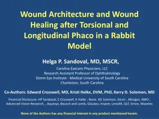
Corneal Epithelial Wound Closure in the Rabbit Model to Compare Besivance TM to Vigamox TM and a Negative Control. B.E. McCarey 1 and P.C. Cockrum 2 Emory University Eye Center, Atlanta, GA 2 Alcon Laboratories, Fort Worth, TX 2

E N D
Corneal Epithelial Wound Closure in the Rabbit Model to Compare Besivance TM to Vigamox TM and a Negative Control B.E. McCarey 1 and P.C. Cockrum 2 Emory University Eye Center, Atlanta, GA 2 Alcon Laboratories, Fort Worth, TX 2 McCarey has received research funds from Alcon Laboratories. Cockrum is an employee of Alcon Laboratories.
Introduction The corneal epithelium is a functional barrier between the tear environment and intraocular environment. An intact epithelium may have a role in preventing stromal haze after laser assisted in situ keratomileusis. As, refractive and cataract procedures increase in number, the effects of medications on corneal epithelial wound healing are of interest. Fluoroquinolones, moxifloxacin and besifloxacin, have been used to treat bacterial infections of the eyes. They are potent against a wide range of bacterial pathogens due to their unique dual binding mechanism of action in gram-positive and gram-negative organisms by inhibition of topiosomerase II (DNA gyrase) and topiosomerase IV.
Purpose The corneal epithelial wound closures rates were compared following topical applications of moxifloxacin and besifloxacin in the rabbit model.
Methods Test Solutions: Besivance TM (Bausch & Lomb) contains 0.6% besifloxacin with 0.01% benzalkonium chloride Vigamox TM (Alcon Laboratories) contains 0.5% moxifloxacin with no preservative BSS TM (Alcon Laboratories) a sterile balanced salt solution with no preservative Treatment: Three treatment groups of 10 rabbits per treatment with bilateral topical eye drops: Group 1: BSS TM and Besivance TM Group 2: BSS TM and Vigamox TM Group 3: Besivance TM and Vigamox TM Test solutions were applied as a single drop QID one day prior to the corneal epithelial wound and QID starting immediately post-wounding.
Methods Animal Model: NZW Rabbit 2.9 to 3.3 kg body weight Surgery: Anesthesia with ketamine and xylazine (30 mg/kg and 6 mg/kg) given IM A Beaver TM blade #5700 was used to scrape away the corneal epithelial cell layer within a 5.0 mm trephine mark. A smooth dull luster from the stromal bed confirmed complete epithelial cell layer removal.
Data Collection A Topcon Photographic slit lamp with a digital camera was used to record the epithelial wounds which were stained with 1.5 µl of 0.5% fluorescein in BSS TM The surface area of the wound was determined with Sigma Scan Pro (Jandel Scientific). The radius of the equivalent area was calculated and plotted relative to post-surgical time. The lineal wound closure rate was determined as described by Crossen et al 1986 (Invest. Ophthalmol. & Vis. Sci. 27:464-473, 1986).
Results Figure 1: The slit lamp photographs are an examples of the corneal epithelial wound stained with sodium fluorescein and treated with Vigamox. The large defect (Figure A) approached closure during the 28 hour follow up (Figure B). Figure 2: An example of the corneal epithelial wound radius closure plot for a rabbit treated with BSS and Vigamox.
Results Figure 3: The rabbit epithelia cell layer closure rates are plotted for the paired eyes from each treatment group. The standard deviation bars for the means are included on the graph. Table 1: Statistical analysis (paired t-test) of the Mean Wound Closure Radii for the Treatment Groups (* statistical difference between treatment groups)
Results Table 2: The Mean Wound Closure Radii for the Treatment Groups (unpaired) Table 3: Statistical analysis (unpaired t-test) of the Mean Wound Closure Radii for the Treatment Groups (* statistical difference between treatment groups) Figure 4: A bar graph representation of the Corneal Epithelial Wound Radius Closure Rates.
Conclusion • The paired t-test of the mean wound closure rates show no statistical difference between the BSS treated eyes and the paired Vigamox treated eyes. However, there is a statistical difference with BSS and Besivance paired eyes and Vigamox and Besivance paired eyes (Table 1). • Regrouping the data as unpaired treatments for epithelial wound closure rates show (Table 3): • BSS (85 µm/hr) to be statistically equivalent to Vigamox (80 µm/hr), • Besivance (68 µm/hr) is statistically slower than BSS (80 µm/hr), and • Vigamox (80 µm/hr) is statistically faster than Besivance (68 µm/hr).