Download
1 / 46
490 likes | 877 Views
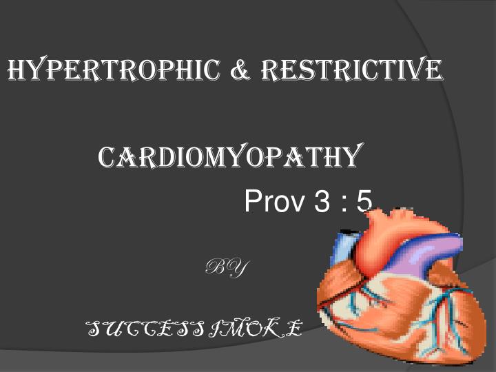
HYPERTROPHIC & RESTRICTIVE CARDIOMYOPATHY Prov 3 : 5 BY SUCCESS IMOKE. INTRODUCTION EPIDEMIOLOGY ETIOLOGY AND RISK FACTORS PATHOPHYSIOLOGY/MORPHOLOGY CLINICAL PRESENTATION DIAGNOSIS TREATMENT. Cardiomyopathy is a disease of the heart muscle

E N D
HYPERTROPHIC & RESTRICTIVE CARDIOMYOPATHY Prov 3 : 5 BY SUCCESS IMOKE
INTRODUCTION EPIDEMIOLOGY ETIOLOGY AND RISK FACTORS PATHOPHYSIOLOGY/MORPHOLOGY CLINICAL PRESENTATION DIAGNOSIS TREATMENT
Cardiomyopathy is a disease of the heart muscle in which the heart loses its ability to pump blood effectively. The heart muscle becomes enlarged or abnormally thick or rigid. In rare cases, the muscle tissue in the heart is replaced with scar tissue.
As cardiomyopathy progresses the heart becomes weaker and less able to pump blood through the body which leads to: Heart failure Arrhythmias Systemic and pulmonary edema Endocarditis
Functional classification • Dilated cardiomyopathy • Hypertrophic cardiomyopathy • Restrictive cardiomyopathy • Arrhthymogenic Right Ventricular disease(ARVD)
HYPERTROPHIC CARDIOMYOPATHY • The irregular growth/thickening of the heart muscle, it affects the main blood pumping muscle • The thicker it becomes, the heart stiffens and the pumping chamber shrinks • The thickening interferes with the hearts capability to deliver blood to the body. myocardium
EPIDEMIOLOGY • Hypertrophic cardiomyopathy affects an estimated 600,000 to 1.5 million Americans, or 1 in 500 people or 0.20%. • It is more prevalent than multiple sclerosis, which affects 1 in 700 people. • 544,000 people in USA have HCM. • About 50% of patients with hypertrophic cardiomyopathy will have a first-degree relative who also has HCM
ETIOLOGY • Genetic (IHSS) • Chronic hypertension • Aging • Idiopathic • There is 5% annual risk of sudden death in all patients who develop HCM
There are two pattern of inheritance: • 60-70% of people develop hypertrophic cardiomyopathy by inheriting a gene that predisposes them to the disease (IHSS) • The second is sporadic
RISK FACTORS • Drop in blood pressure during exercise • Family history of cardiac arrest • History of unexplained syncope • Life-threatening heart rhythms • Severe heart muscle thickness • History of arrhythmia with tachycardia
TYPES OF HCM Obstructive type - the septum thickens and bulges into the left ventricle blocks the flow of blood into the aorta the ventricle must work much harder to pump blood - symptoms can include chest pain, dizziness, DOE, syncope. - can also affect the mitral valve, causing regurgitation.
Non-obstructive type - the entire ventricle may become thicker (symmetric ventricular hypertrophy) or it may happen only at the base of the heart or apex. The right ventricle also may be affected.
Sub aortic Midventricular Apical
HISTOLOGYMuscle cell disarrayInterspersed between areas of hypertrophy Cells are shorter and wider Abnormal orientation of myofibrilsAvg degree of disarray is 30% ( >5% is diagnostic of HOCM) Forceful LV contraction Rapid early ventricular emptying Reduced compliance and impaired relaxation
PATHOPHYSIOLOGY Left ventricular hypertrophy (thick ventricular wall) ventricular chamber size hold less blood CO pressure in the ventricles and lungs changes in the cardiac muscles interfere with the heart's electrical signals, leading to arrhythmias sudden cardiac arrest
CLINICAL PRESENTATION Degree of symptoms not related to level of gradient, but if Gradient >100 mmHg – definitely symptomatic Symptoms can be Latent ( provocable)/ labile/ Persistent M/c/c of sudden death of young athletes in 1st attack. Can present at any age, avg 3rd – 4th decade
SYMPTOMS • Dyspnea on exertion (90%) - Reduced diastolic relaxation & LV filling • Angina (70-80%) - Inadequate myocardial perfusion • Syncope (20%) - Vasovagal attack/ transient arrhythmias • Palpitation (10%) - AF - AF onset - Loss of atrial contribution in LV filling - Rapid rate - 50 % pts of AF manifests as systemic embolisation
SYMPTOMS CONTD • Sudden Cardiac death At later stages Sever progressive heart failure PND/ orthopnea Pulmonary edema Ascites
Physical Examination Cardinal signs • S4 gallop • Palpable LA contractions • Double or triple Apical Impulse • Systolic ejection murmur - Late onset, b/w Lt sternal border & apex
Dynamic maneuvers Murmur intensity increases ( reduced LV size ---- raises level of obstruction) --Reduced Preload – Valsalva/ standing/ tachycardia --Reduced after load -- Vasodilators --Raised contractility – ExerciseMurmur intensity decreases --Increased preload -- squatting, hand grip --Increased after load --Reduced contractility – B blockers
Diagnostic Evaluation • Family history • ECG • X ray • Echocardiogram
Electrocardiogram LV strain pattern LBBB/ RBBB / Lt ant hemiblock
X rayCardiomegalyLA enlargementSmall aortaPulmonary edema
Differential Dx • Aortic stenosis • (concentric hypertrophy, decreased murmur on valsalva, dynamic cath differences) • Restrictive CM • Athlete’s Heart • CAD
Treatment • Medical therapy • Device therapy • Surgical septalmyectomy • Alcohol septal ablation • Transplantation
ACC Consensus Document. J Am Coll Cardiol. 2003;42(9):1693.
Medical Therapy • Beta-blockers • Increase ventricular diastolic filling/relaxation • Decrease myocardial oxygen consumption • Have not been shown to reduce the incidence of SCD • Verapamil • Augments ventricular diastolic filling/relaxation • Disopyramide • Used in combination with beta-blocker • Negative Inotrops • Anticoagulation
Surgical management Septalmyectomy Trans aortic / Left ventriculotomyExtended septalmyectomy mitral valve replacement
Surgical SeptalMyectomy Nishimura RA et al. NEJM. 2010. 350(13):1320.
RESTRICTIVE CARDIOMYOPATHY • Hallmark: abnormal diastolic function • Rigid ventricular wall with impaired ventricular filling ,contractile functions are normal • Much less common than DCM or HCM • Characterized by primary disease in ventricular compliance resulting in impaired ventricular filling during diastole
RESTRICTIVE CARDIOMYPATHY • Idiopathic or systemic myocardial disease characterized by: • Impaired ventricular filling • Elevated diastolic pressures • Normal or reduced diastolic volume of ventricle(s) • Normal/near normal systolic function until advanced stages
ETIOLOGY • Primary---idiopathic • Associated with – • CVD • Amyloidosis • Hemochromatosis • Sarcoidosis • Metastatic tumors • Radiation fibrosis
MORPHOLOGY • Ventricles are of normal size • Cavities are not dilated • Myocardium is firm and non compliant • Biatrial dilatation is common • Patchy/diffuse interstitial fibrosis
RISK FACTORS • Having a family history of cardiomyopathy , heart failure, or sudden cardiac death • Having a disease or condition that can lead to cardiomyopathy, such as: • Coronary artery disease • A previous heart attack • Myocarditis • Diseases that can damage the heart (for example, hemochromatosis, sarcoidosis, or amyloidosis) • Long-term alcoholism • Long-term high blood pressure • Diabetes and other metabolic diseases
SIGNS AND SYMPTOMS Some have no symptoms in the early stages of the disease as cardiomyopathy progresses and the heart weakens, Signs and symptoms of heart failure usually appear: Tiredness Weakness Dyspnea on exertion or at rest Ascities Pedal edema Other signs and symptoms: dizziness, lightheadedness, syncope during exercise, abnormal heart rhythms, murmurs
Clinical manifestations • Symptoms of right and left heart failure • Echo-Doppler • Abnormal mitral inflow pattern • Almost invariably progresses to congestive • heart failure,10% survive for 10 yrs
Amyloidosis • Cardiac enlargement without ventricular dilatation • Ventricular walls are thickened and rubbery • Amyloid deposition is most prominent in interstitial, perivascular and endocardial regions
Endomyocardial diseases • Endomyocardial fibrosis • Loefflersendomyocarditis
Hemochromatosis Sarcoidosis
DIAGNOSIS • Medical history • Physical exam • ECG, chest X-ray • Echocardiogram • Exercise stress test, cardiac catheterization, CT scan, and MRI.
TREATMENT • Treat underlying condition • Diuretics, which remove excess fluid and sodium from the body. • ACE inhibitors, which lower blood pressure and reduce stress on the heart. • Beta-blockers • Calcium channel blockers • Corticosteroid
REFERENCESElliott P, Andersson B, Arbustini E, et al; Classification of the cardiomyopathies: a position statement from the European Eur Heart J. 2008 Jan;29(2):270-6. Epub 2007 Oct 4.Elias TC, Partridge JS, McCorkell SA, et al; A patient with hypertrophic cardiomyopathy undergoing non-cardiac surgery. BMJ. 2013 Nov 25;347:f6910. doi: 10.1136/bmj.f6910.Maron BJ, Maron MS; Hypertrophic cardiomyopathy. Lancet. 2013 Jan 19;381(9862):242-55. doi: 10.1016/S0140-6736(12)60397-3. Epub 2012 Aug 6.Maron MS; Clinical utility of cardiovascular magnetic resonance in hypertrophic cardiomyopathy. J CardiovascMagnReson. 2012 Feb 1;14:13. doi: 10.1186/1532-429X-14-13.Coats CJ, Elliott PM; Genetic biomarkers in hypertrophic cardiomyopathy. Biomark Med. 2013 Aug;7(4):505-16. doi: 10.2217/bmm.13.79.Enriquez AD, Goldman ME; Management of Hypertrophic Cardiomyopathy. Ann Glob Health. 2014 January - February;80(1):35-45. doi: 10.1016/j.aogh.2013.12.004. Epub 2013 Dec 25.Guttmann OP, Rahman MS, O'Mahony C, et al; Atrial fibrillation and thromboembolism in patients with hypertrophic cardiomyopathy: systematic review. Heart. 2014 Mar;100(6):465-72. doi: 10.1136/heartjnl-2013-304276. Epub 2013 Sep 7.Said SM, Dearani JA, Ommen SR, et al; Surgical treatment of hypertrophic cardiomyopathy. Expert Rev CardiovascTher. 2013 May;11(5):617-27. doi: 10.1586/erc.13.46.Gersh BJ, Maron BJ, Bonow RO, et al; 2011 ACCF/AHA guideline for the diagnosis and treatment of hypertrophic cardiomyopathy: a report of the American College of Cardiology