Download
1 / 22
220 likes | 364 Views
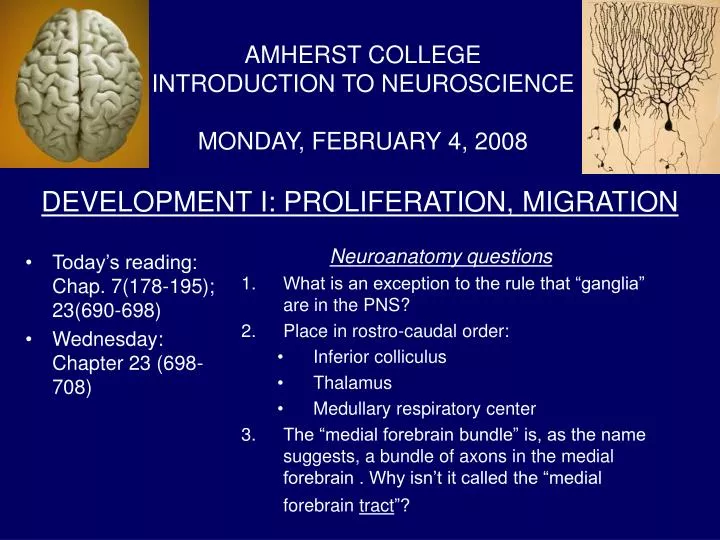
DEVELOPMENT I: PROLIFERATION, MIGRATION. AMHERST COLLEGE INTRODUCTION TO NEUROSCIENCE MONDAY, FEBRUARY 4, 2008. Today’s reading: Chap. 7(178-195); 23(690-698) Wednesday: Chapter 23 (698-708). Neuroanatomy questions What is an exception to the rule that “ganglia” are in the PNS?

E N D
DEVELOPMENT I: PROLIFERATION, MIGRATION AMHERST COLLEGE INTRODUCTION TO NEUROSCIENCEMONDAY, FEBRUARY 4, 2008 • Today’s reading: Chap. 7(178-195); 23(690-698) • Wednesday: Chapter 23 (698-708) Neuroanatomy questions • What is an exception to the rule that “ganglia” are in the PNS? • Place in rostro-caudal order: • Inferior colliculus • Thalamus • Medullary respiratory center • The “medial forebrain bundle” is, as the name suggests, a bundle of axons in the medial forebrain . Why isn’t it called the “medial forebrain tract”?
Tomorrow - 7:00 AM to 8:00 PM Bangs Community Center (Behind Panda East & Rao’s)
Proliferation Fertilized egg Whole organism (single cell) 1011 neurons → Question: how many rounds of cell division? 2x2x2x2x…. = 10x10x10x….x10 Log10(2)=3.3, i.e.10 = 23.3, so 1011=(23.3)11= 236
Neural crest: -Dorsal root ganglia -Autonomic ganglia -PNS myelin CNS Notochord Early vertebrate development Neural groove →neural tube. Vertebrate CNS = hollow organ Fig. 7.8
Vertebrate brain development: 3 subdivisions Anterior neural tube → 3 brain ‘vesicles’ → Basal Line of Thalamus ganglia “secondary fusion” Fig. 7.9, 7.10, 7.13
Development of the retina Retinal ganglion cells develop after the optic cup is formed. Their axons grow from the retina to the diencephalon and midbrain. Is “optic nerve” a consistent name for the bundle of axons connecting the retina to the rest of the brain? Is “ganglion” cell a consistent name for neurons in the retina? Fig. 7.10,11
Features of proliferation • Cell divisions occur in ventricular zone • Nucleus moves to marginal zone for “S” phase (synthesizing DNA for next division) • Cell becomes postmitotic (has its “birthday”) after horizontal cleavage Fig. 22.2
1. Technique: Autoradiography 3H = tritium, hydrogen atom with 2 extra neutrons; it undergoes radioactive decay 3H -thymidine (a nucleotide that is one component of the genetic material, DNA) is used to study cell “birthdays” (i.e. date of final cell division) 3H-proline, an amino acid that is one component of proteins, is used to trace neuronal pathways.
Analysis of cell birthdays Label monkey fetus with 3H-thymidine at E33 or E56 Wait until animal matures, then perform autoradiography Pia Conclusion: Postmitotic neurons migrate towards the pia, migrating past neurons that migrated previously. Exposed to label at E56 Exposed to label at E33 Ventricle
Technical question: When a monkey fetus is exposed to 3H-thymidine at embryonic day 33, shouldn’t all dividing cells take up the label, including cells whose progeny will be dividing later, e.g. at day 56? In that case, why is there only a narrow band of label in the animals exposed at day 33 – why doesn’t it extend all the way up to the pia? Exposed at E33 Exposed at E56
Features of mammalian cortical development • Dividing neuroblasts proliferate in the ventricular zone • At some point (before birth in mammals) cells stop dividing – i.e. they have their “birthday,” except for a very few neuronal stem cells • They immediately migrate towards the marginal zone, following radial glial cells • The order of migration is “inside out,” i.e. newly born neurons migrate past previously migrated ones to a point nearer the pia • Large neurons are born earlier than small neurons • Glial cells retain the capability of dividing throughout the life of the animal
Like DNA Fluorescent Neurons and the “cell cycle” • Are neurons able to divide, or are they “post-mitotic?” • (Mitosis = cell division; post-mitotic = can no longer divide) • Look for “mitotic figures” in slices of brain tissue • But, there’s a problem… • Modern methods • Autoradiography • Bromodeoxyuridine (BRDU)
2. BRDU + specific neuron label Green=BRDU Red=neuron-specific stain
Neuronal differentiation: Control of gene expression membrane membrane nucleus nucleus cytoplasm cytoplasm
membrane nucleus cytoplasm
Introduction to cell death • 2 – 3 times overproduction of cells during development • Occurs in many parts of the body • Two distinct kinds: • Programmed (apoptotic, “suicide”) • Necrotic (“murder”) • In invertebrates, specific identified cell always dies at a particular time • Cell death program can be activated in adults, e.g. neurodegenerative diseases