Download

1 / 51
510 likes | 577 Views
Pressure and Resistance. Fluid Recycling Water continuously moves out of capillaries, and back into bloodstream via the lymphoid system and serves to Ensure constant plasma and interstitial fluid communication Accelerate distribution of nutrients, hormones, and dissolved gases through tissues

E N D
Pressure and Resistance • Fluid Recycling • Water continuously moves out of capillaries, and back into bloodstream via the lymphoid system and serves to • Ensure constant plasma and interstitial fluid communication • Accelerate distribution of nutrients, hormones, and dissolved gases through tissues • Transport insoluble lipids and tissue proteins that cannot cross capillary walls • Flush bacterial toxins and chemicals to immune system tissues
Pressure and Resistance • Capillary Dynamics • Hemorrhaging • Reduces CHP and NFP • Increases reabsorption of interstitial fluid (recall of fluids) • Dehydration • Increases BCOP • Accelerates reabsorption • Increase in CHP or BCOP • Fluid moves out of blood • Builds up in peripheral tissues (edema)
Cardiovascular Regulation • Tissue Perfusion • Blood flow through the tissues • Carries O2 and nutrients to tissues and organs • Carries CO2 and wastes away • Is affected by • Cardiac output • Peripheral resistance • Blood pressure
Cardiovascular Regulation • Cardiovascular regulation changes blood flow to a specific area • At an appropriate time • In the right area • Without changing blood pressure and blood flow to vital organs
Cardiovascular Regulation Figure 19–13 Short-Term and Long-Term Cardiovascular Responses
Cardiovascular Regulation • Controlling Cardiac Output and Blood Pressure • Autoregulation • Causes immediate, localized homeostatic adjustments • Neural mechanisms • Respond quickly to changes at specific sites • Endocrine mechanisms • Direct long-term changes
Cardiovascular Regulation • Autoregulation of Blood Flow within Tissues • Adjusted by peripheral resistance while cardiac output stays the same • Local vasodilators: • accelerate blood flow at tissue level • low O2 or high CO2 levels • low pH (acids) • nitric oxide (NO) • high K+ or H+ concentrations • chemicals released by inflammation (histamine) • elevated local temperature
Cardiovascular Regulation • Autoregulation of Blood Flow within Tissues • Adjusted by peripheral resistance while cardiac output stays the same • Local vasoconstrictors: • examples include prostaglandins and thromboxanes • released by damaged tissues • constrict precapillary sphincters • affect a single capillary bed
Cardiovascular Regulation • Neural Mechanisms • Cardiovascular (CV) centers of the Medulla Oblongata • Cardiac centers: • cardioacceleratory center: increases cardiac output • cardioinhibitory center: reduces cardiac output • Vasomotor center: • vasoconstriction • controlled by adrenergic nerves (NE) • stimulates smooth muscle contraction in arteriole walls • vasodilation: • controlled by cholinergic nerves (NO) • relaxes smooth muscle
Cardiovascular Regulation • Vasomotor Tone • Produced by constant action of sympathetic vasoconstrictor nerves
Cardiovascular Regulation • Reflex Control of Cardiovascular Function • Cardiovascular centers monitor arterial blood • Baroreceptor reflexes: • respond to changes in blood pressure • Chemoreceptor reflexes: • respond to changes in chemical composition, particularly pH and dissolved gases
Cardiovascular Regulation • Baroreceptor Reflexes • Stretch receptors in walls of • Carotid sinuses: maintain blood flow to brain • Aortic sinuses: monitor start of systemic circuit • Right atrium: monitors end of systemic circuit • When blood pressure rises, CV centers • Decrease cardiac output • Cause peripheral vasodilation • When blood pressure falls, CV centers • Increase cardiac output • Cause peripheral vasoconstriction
Cardiovascular Regulation Figure 19–14 Baroreceptor Reflexes of the Carotid and Aortic Sinuses
Cardiovascular Regulation • Chemoreceptor Reflexes • Peripheral chemoreceptors in carotid bodies and aortic bodies monitor blood • Central chemoreceptors below medulla oblongata • Monitor cerebrospinal fluid • Control respiratory function • Control blood flow to brain
Cardiovascular Regulation • Chemoreceptor Reflexes • Changes in pH, O2, and CO2 concentrations • Produced by coordinating cardiovascular and respiratory activities
Cardiovascular Regulation Figure 19–15 The Chemoreceptor Reflexes
Cardiovascular Regulation • CNS Activities and the Cardiovascular Centers • Thought processes and emotional states can elevate blood pressure by cardiac stimulation and vasoconstriction
Cardiovascular Regulation • Hormones and Cardiovascular Regulation • Hormones have short-term and long-term effects on cardiovascular regulation • For example, E and NE from suprarenal medullae stimulate cardiac output and peripheral vasoconstriction
Cardiovascular Regulation • Antidiuretic Hormone (ADH) • Released by neurohypophysis (posterior lobe of pituitary) • Elevates blood pressure • Reduces water loss at kidneys • ADH responds to • Low blood volume • High plasma osmotic concentration • Circulating angiotensin II
Cardiovascular Regulation • Angiotensin II • Responds to fall in renal blood pressure • Stimulates • Aldosterone production • ADH production • Thirst • Cardiac output • Peripheral vasoconstriction
Cardiovascular Regulation • Erythropoietin (EPO) • Released at kidneys • Responds to low blood pressure, low O2 content in blood • Stimulates red blood cell production
Cardiovascular Regulation • Natriuretic Peptides • Atrial natriuretic peptide (ANP) • Produced by cells in right atrium • Brain natriuretic peptide (BNP) • Produced by ventricular muscle cells • Respond to excessive diastolic stretching • Lower blood volume and blood pressure • Reduce stress on heart
Cardiovascular Regulation Figure 19–16a The Hormonal Regulation of Blood Pressure and Blood Volume.
Cardiovascular Regulation Figure 19–16b The Hormonal Regulation of Blood Pressure and Blood Volume.
Cardiovascular Adaptation • Blood, heart, and cardiovascular system • Work together as unit • Respond to physical and physiological changes (for example, exercise, blood loss) • Maintains homeostasis
Cardiovascular Adaptation • The Cardiovascular Response to Exercise • Light exercise • Extensive vasodilation occurs: • increasing circulation • Venous return increases: • with muscle contractions • Cardiac output rises: • due to rise in venous return (Frank–Starling principle) and atrial stretching
Cardiovascular Adaptation • The Cardiovascular Response to Exercise • Heavy exercise • Activates sympathetic nervous system • Cardiac output increases to maximum: • about four times resting level • Restricts blood flow to “nonessential” organs (e.g., digestive system) • Redirects blood flow to skeletal muscles, lungs, and heart • Blood supply to brain is unaffected
Cardiovascular Adaptation • Exercise, Cardiovascular Fitness, and Health • Regular moderate exercise • Lowers total blood cholesterol levels • Intense exercise • Can cause severe physiological stress
Cardiovascular Adaptation • The Cardiovascular Response to Hemorrhaging • Entire cardiovascular system adjusts to • Maintain blood pressure • Restore blood volume
Cardiovascular Adaptation • Short-Term Elevation of Blood Pressure • Carotid and aortic reflexes • Increase cardiac output (increasing heart rate) • Cause peripheral vasoconstriction • Sympathetic nervous system • Triggers hypothalamus • Further constricts arterioles • Venoconstriction improves venous return
Cardiovascular Adaptation • Short-Term Elevation of Blood Pressure • Hormonal effects • Increase cardiac output • Increase peripheral vasoconstriction (E, NE, ADH, angiotensin II)
Cardiovascular Adaptation • Shock • Short-term responses compensate after blood losses of up to 20% of total blood volume • Failure to restore blood pressure results in shock Circulatory Shock
Cardiovascular Adaptation • Long-Term Restoration of Blood Volume • Recall of fluids from interstitial spaces • Aldosterone and ADH promote fluid retention and reabsorption • Thirst increases • Erythropoietin stimulates red blood cell production
Cardiovascular Adaptation Figure 19–17 Cardiovascular Responses to Hemorrhaging and Blood Loss
Cardiovascular Adaptation • Vascular Supply to Special Regions • Through organs with separate mechanisms to control blood flow • Brain • Heart • Lungs
Cardiovascular Adaptation • Blood Flow to the Brain • Is top priority • Brain has high oxygen demand • When peripheral vessels constrict, cerebral vessels dilate, normalizing blood flow
Cardiovascular Adaptation • Stroke • Also called cerebrovascular accident (CVA) • Blockage or rupture in a cerebral artery • Stops blood flow
Cardiovascular Adaptation • Blood Flow to the Heart • Through coronary arteries • Oxygen demand increases with activity • Lactic acid and low O2 levels • Dilate coronary vessels • Increase coronary blood flow • Epinephrine • Dilates coronary vessels • Increases heart rate • Strengthens contractions
Cardiovascular Adaptation • Heart Attack • A blockage of coronary blood flow • Can cause • Angina (chest pain) • Tissue damage • Heart failure • Death
Cardiovascular Adaptation • Blood Flow to the Lungs • Regulated by O2 levels in alveoli • High O2 content • Vessels dilate • Low O2 content • Vessels constrict
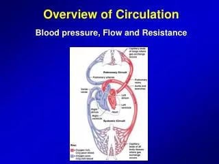
Pulmonary and Systemic Patterns Figure 19–18 A Schematic Overview of the Pattern of Circulation.
The Pulmonary Circuit • Deoxygenated blood arrives at heart from systemic circuit: • Passes through right atrium and right ventricle • Enters pulmonary trunk • At the lungs: • CO2 is removed • O2 is added • Oxygenated blood: • Returns to the heart • Is distributed to systemic circuit
The Pulmonary Circuit • Pulmonary Vessels • Pulmonary arteries • Carry deoxygenated blood • Pulmonary trunk: • branches to left and right pulmonary arteries • Pulmonary arteries: • branch into pulmonary arterioles • Pulmonary arterioles: • branch into capillary networks that surround alveoli
The Pulmonary Circuit • Pulmonary Vessels • Pulmonary veins • Carry oxygenated blood • Capillary networks around alveoli: • join to form venules • Venules: • join to form four pulmonary veins • Pulmonary veins: • empty into left atrium
The Pulmonary Circuit Figure 19–19 The Pulmonary Circuit
The Systemic Circuit • Contains 84% of blood volume • Supplies entire body • Except for pulmonary circuit
The Systemic Circuit • Systemic Arteries • Blood moves from left ventricle • Into ascending aorta • Coronary arteries • Branch from aortic sinus