Download
1 / 30
310 likes | 505 Views
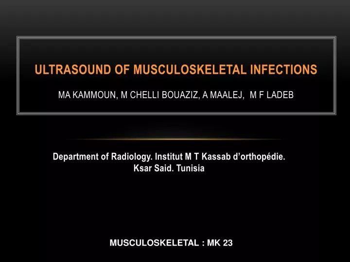
ULTRASOUND OF MUSCULOSKELETAL INFECTIONS MA KAMMOUN, M CHELLI BOUAZIZ, A MAALEJ, M F LADEB. Department of Radiology . Institut M T Kassab d’orthopédie. Ksar Said. Tunisia. MUSCULOSKELETAL : MK 23. INTRODUCTION.

E N D
ULTRASOUND OF MUSCULOSKELETAL INFECTIONSMA KAMMOUN, M CHELLI BOUAZIZ, A MAALEJ, M F LADEB Department of Radiology. Institut M T Kassab d’orthopédie. Ksar Said. Tunisia MUSCULOSKELETAL : MK 23
INTRODUCTION • Musculoskeletal infections are commonly encountered in clinical practice in children and adult patients • Radiographs remain the first imaging modality to perform in these conditions • Ultrasound (US) may be used either as the primary imaging technique or as an adjunct to radiography, computed tomography (CT), magnetic resonance imaging (MRI) and nuclear medicine studies
OSTEOMYELITIS Acute osteomyelitis • Daily US examination allows an early detection of subperiosteal abcess thus indicating surgical treatment (protocol of Tunis). • Clinical and US differential diagnosis is sometimes difficult with sickle cell anemia vaso-occusive crisis and subperiosteal haematoma.
Acute osteomyelitis: subperiosteal abcess of the femur is well assessed with US
Acute osteomyelitis: subperiosteal abcess of the tibia is well assessed with US
Vaso-occlusive crisis. US shows a subperiosteal haematoma of the tibia Vaso-occlusive crisis. US shows a subperiosteal haematoma
OSTEOMYELITIS Chronic osteomyelitis • Soft tissue modifications and /or Juxtacortical collections are assessed with US in acute reactivation of chronic osteomyelitis. • Fistula , soft tissue sequestra and cortical bone modifications are also well assessed with US
* Chronic osteomyelitis reactivation: Juxtacortical abcess with sequestrum
* Chronic osteomyelitis reactivation: Juxtacortical abcess with a fistula.(*) Reactivation of a chronic osteomyelitis. US shows cortical bone irregularities and calcifiactions with a soft tissue abcess
ARTHRITIS • In acute arthritis, US shows a joint effusion with or without synovial thickening and local hyperhemia. • Bone abnormalities such as periosteal new bone formation or perichondral erosions are also well assessed by US
ARTHRITIS • Chronic arthritis may show a similar appearance • A local amyotrophy around the joint may be observed. Several ultrasonographic signs may help to identify specific infections
ARTHRITIS • The importance of synovial thickening and the presence of thin calcifications into the synovium suggests a tuberculous origin whereas a multicystic appearance is characteristic of echinococcosis
Acute knee arthritis. US shows a joint effusion with synovial thickening
Acute arthritis of the elbow. US Shows a joint effusion without synovial thickening
Acute arthritis of the knee. US shows an important synovial thickening with joint effusion and local hyperhemia.
* ** Osteo arthritis of the first metatarsophlangeal joint. Bone abnormalities such as periosteal new bone formation (*) and perichondral bone erosion (**) are easily assessed by US
* Tuberculous arthritis of the knee. Note the importance of the synovial thickening and the fine synovial calcification(*). Echinococcosis of the iliac bone. US shows a characteristic multicystic appearance in the soft tissues.
Infection of the skin and subcutaneous tissue Clinical diagnosis often obvious: Sudden onset of local and general inflammatory closet "orange peel“ Alteration of general state, fever, ganglia INFECTIOUS CELLULITIS
Radiographs: nonspecific Ultrasound: abscessDiffuse thickening of the skin"dissected appearance" of subcutaneous fat lobulesHyperemia at color Doppler CT / MRI:Infiltration of subcutaneous fatHypo T1, T2 Hyper without mass effectEnhancement after contrast injection+ / - Edema of the fascia and adjacent muscles INFECTIOUSCELLULITISIMAGING
* Radiographs showing thikening of sucutaneous fat (*). US: soft tissue thickening and Doppler hyperhaemia soft tissue cellulitis: US shows "dissected appearance" of subcutaneous fat lobules
Pyomyositis: muscle abscess Phlegmon: inflammatory infiltration of the muscle not collected before the collected stage PHLEGMON AND PYOMYOSITIS
Pain Muscle induration History of local trauma: from 22 to 67% A single muscle group is usually affected Careful analysis of adjacent bones and joints to confirm the muscular origin of the infection PHLEGMON AND PYOMYOSITISCLINICAL PRESENTATION
Radiographs : of little use Ultrasound:Increased muscle volumehypoechoic septaEvolution towards the abscess with hypoechoic center + / - standard liquid or thin wallsEchogenic wall + / - thick that may contain calcificationsImaging can guide the puncture PHLEGMON AND PYOMYOSITISIMAGING
Pyomyositis of the thigh: US shows muscle thickening, heterogenous appearance and colour Doppler hyperhemia.
Pyo-myositis in 33 years old man. US shows global thikening of biceps muscle and presence of liquid collection (abcess) into the muscle.
Clinical presentation: local inflammatory syndrome Fever: 40% of cases Germ: staph aureus X-rays + CT: Swelling of the bursa Ultrasound: Thickening of the bursa wall, echogenic content , Doppler hyperemia MRI: staging SEPTIC BURSITIS
Septic bursitis: Ultrasound shows thickening of the subacromial subdeltoid bursa wall with color Doppler hyperemia and fluid collection
Often by inoculation Staph aureus, tuberculosis Fingers and toes flexors Radiographs: eliminate arthritis or osteitis Ultrasound: thickening of tendon sheath + / - effusion, Doppler hyperemia MRI: thickening of tendon sheath, enhancement after contrast injection SEPTIC TENOSYNOVITIS
Tenosynovitis: US shows thickening of the tendon sheath + / - effusion, Doppler hyperhemia Note also the presence of little abcesses into the synovial sheath
CONCLUSION • Ultrasound is very performant in the diagnosis and follow up of musculoskeletal infections. • It allows to: • differentiate infection from tumors or non-infective inflammatory conditions with similar clinical presentation • localize the site and extent of infection • guide drainage or biopsy