Download
1 / 63
690 likes | 878 Views
Dr. Hakan Arslan. LYMPHEDEMA AND TREATMENT. “ Accumulation of abnormal amount of protein rich fluid in the interstitium due to compromised lymphatic system with (near) normal net capillary filtration ”. In United States

E N D
Dr. Hakan Arslan LYMPHEDEMA AND TREATMENT
“Accumulation of abnormal amount of protein rich fluid in the interstitium due to compromised lymphatic system with (near) normal net capillary filtration”
In United States Highest incidence is observed following breast cancer surgery with radiotherapy (10 – 40%).
Worldwide 140-250 million cases of lymphedema are estimated to exist with filariasis as the most common cause
Lymphatic filariasis affects more than 90 million people in the world
According to WHO Lymphatic Filariasis is the 2nd leading cause of permanent & long term disability in the world after leprosy
Micronatomy of lymphatic system • Lymphatic capillaries • Blind ended • Large intercellular & intracellular • fenestrations • Allowing macromolecular influx (1000 kDa) • Collagen fibers attachment on outer surface • Dermal papillae
Microanatomy of lymphatic system • Sub papillary pre-collectors • Sub-dermal collector lymphatics • Epifacial, valved, muscular lymphatics • with lymphangions • Subfascial lymphatics • Interconnections at inguinal, anticubital, • axillary levels
Capillaries Pre-collectors Collectors Deep lymphatic trunk
10% 90% Pathophysiology
Sub dermal fibrosis Dermal thickening Collagen deposition Valvular incompetence Aplasia hypoplasia Hypocon- tractility LYMPH- EDEMA lymphostasis Obstruction Pathophysiology
LYMPHEDEMA Secondary lymphedema Primary lymphedema Congenital Praecox Tarda Etiology of lymphedema
Congenital lymphedema < 1year of age 10-25% of all primary lymphedema Sporadic or familial (Milroy's disease) More common in males Lower extremity is involved 3 times more frequently than the upper extremity 2/3 patients have bilateral lymphedema Aplasia pattern without subcutaneous lymphatic trunks involvement
Lymphedema Precox Evident after birth and before age 35 years Most often arises during puberty 65-80% of all primary lymphedema cases Females are affected 4 times 70% of cases are unilateral, with the left lower extremity being involved Hypoplastic pattern, with the lymphatics reduced in caliber and number
Lymphedema Tarda (Meige disease ) Clinically not evident until 35 years or older Rarest form of primary lymphedema Only 10% of cases Hyperplasic pattern, with tortuous lymphatics increased in caliber and number Absent or incompetent valves
Secondary Lymphedema Most common lymphedema having well recognized causes
Filariasis Commonest cause worldwide Endemic in 72 countries Affecting 5-10% population Africa, India, South America
Wuchereria Bancrofti (90%) Brugia malayi Brugia timori Filariasis
Other causes of Secondary Lymphedema Breast surgery with radiotherapy Primary malignancy Prostate, cervical cancer, malignant melanoma Trauma to lymphatics Surgical excision of lymph nodes
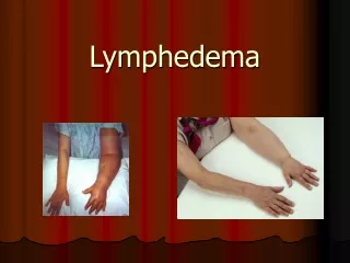
Presentation of lymphedema Age of onset Painless swelling Presence or absence of family history Coexistent pathology
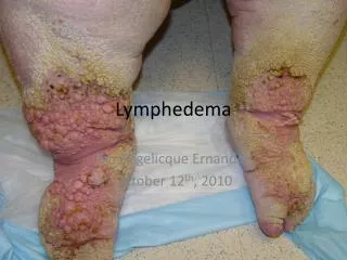
Presentation of lymphedema Characteristically foot involvement Ankle contours are lost with infilling of the submalleolar depressions Buffalo hump on foot dorsum Square shaped toes Stemmer’s sign
Skin changes Chronic eczema Dermatophytosis Fissuring Verrucae Ulcerations Stewart Treves syndrome
III Irreversible skin changes, fibrosis, papillae I Pitting edema, Subsides with elevation II Non pitting edema Not relieved with elevation 0 Histological abnormalities Not clinical evident Brunner Classification
Investigations Infrequently required to establish the diagnosis To determine residual lymphatic function To establish treatment preferences To evaluate therapy
Contrast Lymphangiography Was gold standard for mapping Damages the normal lymphatic channels due to inflammation Very painful procedure and needs GA
Isotope Lymphoscintigraphy Replaced the earlier Technetium labeled antimony sulphide
MRI Scan An indication for CT scan or MRI is suspicion of malignancy, for which these tests offer the most information
Differential diagnosis Congestive heart failure Liver and renal failure Deep vein thrombosis Venous insufficiency Lipedema (usually sparing the feet) Idiopathic edema Hypoalbuminemia Vascular malformations
TREATMENT Conservative Surgical
Conservative Medication Physical
Complex Lymphedema Therapy (CLT) • Manual lymphatic drainage (MLD)* • (massage to make the flow to normal lymphatics) • Low stretch bandaging • (to prevent re-accumulation) *Vodder and/or Leduc techniques
Intermittent pneumatic pump compression therapy Effectively milking the lymph from the extremity Compression garment To help prevent return of fluid
Skin care (Examine, dry, moisturizers) Exercises
Psychological support & occupational therapy
Antiparasitic agents Diethylecarbimazole 6mg/kg single dose or 1-3wk (Don’t use in pregnancy, infants, elderly) Ivermectin (400mcg/kg/d) Tetracycline Doxycycline (100mg/day for 6-8 wks)