Download
1 / 42
470 likes | 1.14k Views
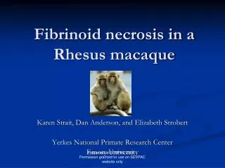
Fibrinoid Necrosis. Fibrinoid necrosis. So called “ fibrinoid degeneration ” in old textbooks of pathology

E N D
Fibrinoid necrosis • So called “fibrinoid degeneration” in old textbooks of pathology • It appears as strongly eosinophilic, reflexible small granules, pieces or amorphous materials, in the wall of blood vessel or in connective tissue. The original structure is destroyed. • It can be seen in rheumatic fever, allergic vasculitis and other immune complex injury (type III hypersensitivity)
Fibrinoid necrosis in an Aschoff nodule from a patient with rheumatic myocarditis.
Sequelae of necrosis • Acute inflammation (autolysis and heterolysis) • Absorption and resolution (break down) • Cyst and abscess • Fall off from a hollow organ • Ulceration and cavity (erosion) • Fistula and sinus • Organization • Calcification and encapsulation • Repair and regeneration
Sequelae of necrosis • Cyst: A closed space contained fluid behind the resolution and absorption of necrotic tissue • Abscess:A localized collection of pus in part of the body, formed by tissue disintegration and surrounded by an inflamed area. • Ulcer: A lesion after loss of a part of tissue in surface of body or in a hollow organ. • Erosion: Loss of only the epithelium of mucosa.
Sequelae of necrosis • Cavity: an empty space connected outside of body through a physiologic channel. • Fistula: An abnormal duct or passage resulting from injury, disease, or a congenital disorderthat connects a hollow organ to the body surface or to another hollow organ. • Sinus: An abnormal duct leading from a necrotic tissue to body surface. • Organization: A process of replacement of necrotic tissue, thrombi, foreign bodies and inflammatory exudate through ingrowth of granulation tissue.
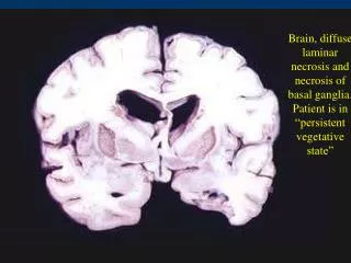
Grossly, the cerebral infarction at the upper left here demonstrates liquefactive necrosis. Eventually, the removal of the dead tissue leaves behind a cyst.
慢性胃溃疡 Gastric ulceration
气管食管瘘 An esophagus-trachea fistula in patient with esophageal carcinoma.
Organization of a old heart infarct. The scars localize In the anterior and lateral walls of left ventricle. 陈旧性心肌梗死机化后瘢痕形成
Apoptosis-Programmed cell death • Apoptosis: a falling away from, like yellow leaves fall away from tree in autumn • Concept: apoptosis is considered as death of single cells in living body which is characteristic by formation of apoptotic bodies and without inflammatory reaction in surrounding tissue. • Cell suicide apoptosis • Homicide necrosis
Apoptosis-Programmed cell death Significance of apoptosis • The remodeling of embryonal tissue • Hormone dependent physiologic and pathologic involution of the • Endometrium during menstrual cycle • Atrophy of testis in old male • Cell depletion of normal intestinal crypt epithelium and tumor cells • Negative selection of immune cells (T cells) in thymus • Cell depletion by TP53 molecules
Different from necrosis, apoptosis is a death of single cell
Morphology of apoptosis • Shrinkage of single cell with eosinophilic cytoplasm and condensed chromatin around the nuclear membrane • Divided into several round or oval globelets with strongly eosinophilic cytoplasm and a part of condensed chromatin (apoptotic bodies) • Phagocytosed by surrounding normal cells or macrophages
Apoptosis is individual cell death, not necrosis (death of large numbers of cells). In this example, liver cells are dying individually (arrows) from injury by viral hepatitis. The shrank cells with dense nuclei are called acidophilic change. Smaller globelets are apoptotic bodies (Councilman’s bodies)
Ultrastructural changes of apoptosis cells: shrinkage of cells → Dried all organelle → peripheral condensed chromatin under the nuclear membrane→well limited smaller dense chromatin plaques → divided into several apoptotic bodies
Ladder pattern Molecular mechanism of apoptosis: DNA fragmentation is from the linker regions of nucleosomes.
The sequential ultrastructural changes in necrosis (left) and apoptosis (right).
Simplified features of coagulative necrosis and apoptosis coagulative necrosis apoptosis Stimuli hypoxia, toxins physiologic and pathologic factors Histologic cellular swelling single cells changes coagulation necrosis chromatin condensation disruption of organelles apoptotic bodies DNA breakdown random, diffuse internucleosomal Mechanisms ATP depletion gene activated membrane injury endonucleased free radical damage Tissue reaction inflammation no inflammation phagocytosis
Cellular aging “We grow too soon old and too late smart.”
Cellualr aging • Cellular aging is the basis of body aging • Aging cells with • Decreased function • Accumulation of metabolic products (brown atrophy-lipofuscin deposition, so called “wear and tear” pigments) • Eventually death through apoptosis, the aged organ appears as atrophy
The yellow-brown granular pigment is lipofuscin (lipochrome) which accumulates over time in cells (particularly liver and heart) as a result of "wear and tear" with aging.
Lipofuscin in the myocardial fiber under the EM: the high electronic density materials are residual bodies (arrow).
Cellualr aging: Why? The mechanism of aging is not clear yet. • Evidences • The normal human fibroblasts in culture have about 50 doubling of span-life • Fibroblasts from neonates: 65 doubling • Why the cell knows their number of divisions? • Telomere shortening theory • Clock gene theory (aging clock)
Telomere shortening • After each doubling, the cell lost a short piece of DNA in the end of chromosomes, so fidelity of DNA replication in the daughter cells can not be sure. • In the normal cells there is a special mechanism to protect the fidelity by using a repeated nontrancribed DNA sequence (TTAGGG)---telomere---as the ends of chromosomes.
Telomere shortening • After each doubling, the telomere has been cut a little to protect the trancribed DNA, so the fidelity of the replication is ensured. • In somatic cells the cell doubling does not continue after multiple cell division, so the cells are aged.
Telomere shortening • But in germ cells, stem cells and neoplastic cells the length of telomere can restore after division by the activation of telomerase. So these cells can continuously replicate • Activation of telomerase is thought of a reason of carcinogenesis.
Clock genes • There are some genes which control the life time in some worms (Caenorhabditis elegans: clk-1 gene mutanted worms have only 50% lifespan compared with normal) • Wear and tear theories • Free radicals play an important role in cell aging • DNA repair mechanism is limited
Summary of cellular aging • Cellular aging is the basis of body aging • Cellular aging involves programmed aging (life timer) and environmental injury (free radicals). • The mechanism of aging can be the highlight of the study of tumorigenesis. • “Long live forever” is impossible.