Download
1 / 23
240 likes | 457 Views
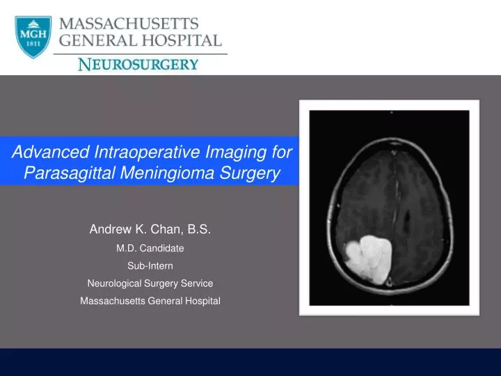
Advanced Intraoperative Imaging for Parasagittal Meningioma Surgery. Andrew K. Chan, B.S. M.D. Candidate Sub-Intern Neurological Surgery Service Massachusetts General Hospital. Case Presentation. HPI 48 year old woman Intermittent, dull, bilateral frontal h eadache x 5 weeks

E N D
Advanced Intraoperative Imaging for Parasagittal Meningioma Surgery Andrew K. Chan, B.S. M.D. Candidate Sub-Intern Neurological Surgery Service Massachusetts General Hospital
Case Presentation • HPI • 48 year old woman • Intermittent, dull, bilateral frontal headache x 5 weeks • Worsened the past 4 days, now persistent, n/v, difficulty focusing • Exam • Left Pronator Drift, 4+/5 Left Upper Extremity Strength
Right Parietal Craniotomy for Resection • Left-lateral position, with head down 60 degrees • Monitoring / Imaging: • Central Venous Catheter • Pre-Cordial Doppler • Intraoperative CT scan for BrainLab frameless stereotaxy • Microscope • Removed tumor nearsinus, and removed medial portion of tumor above a draining vein, that was preserved • Frozen Pathology: Meningioma w/o atypical features
Convexity Meningiomas • 391 convexity meningiomas • 60.1 years (19 – 92 years) • WHO I, II, III (90.3, 5.6, 4.1%) • Median Follow-up: 7.1 years (0.0 – 20.9 years) • 1-, 5-, 10-year survival 96%, 89%, 78% • Overall survival associated with age, sex, WHO grade, Simpson grade • 1-, 5-, 10-year retreatment-free survival 99%, 94%, 90% • Retreatment-free survival associated with WHO grade, Simpson grade Hasseleild et al. 2012, J Neurosurg
Convexity Meningiomas 4.9x 13.2x Hasseleild et al. 2012, J Neurosurg
Eclipse Sign Diminishment of Eclipse Sign Ueba et al. 2013, J Neurosurg
Intraoperative Guidance: Extent of Resection Kim et al. 2011,ActaNeurochir
Intraoperative Guidance: Extent of Resection d’Avella et al. 2013,ActaNeurochir
Conclusions • Aids in the real time, in situ visualization of… • Dural venous sinuses • Cortical arteries and veins • Dural attachment • Select surgical scenarios • Close to major vessels • Approaching highly vascularized tumors • Safe, non-invasive, inexpensive • Future, large series to assess clinical impact
Thank You • Attendings • Residents • Staff • Co-Sub Is • Guy M. McKhann II, MD • Sameer A. Sheth, MD, PhD
Cost Comparison: Transsphenoidal Surgery Eboli et al. 2011, J Neurosurg
Brainlab • Cost • $ 225,000 Watkins et al. 2010 Open Orthop J