Download
1 / 38
450 likes | 907 Views
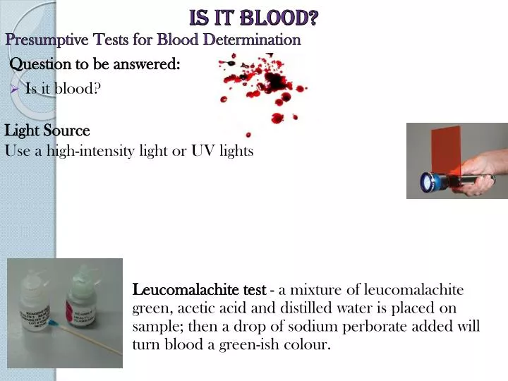
Is it blood? . Presumptive Tests for Blood Determination. Question to be answered: Is it blood?. Light Source Use a high-intensity light or UV lights.

E N D
Is it blood? Presumptive Tests for Blood Determination Question to be answered: • Is it blood? Light Source Use a high-intensity light or UV lights Leucomalachite test - a mixture of leucomalachite green, acetic acid and distilled water is placed on sample; then a drop of sodium perborate added will turn blood a green-ish colour.
Is It blood? • Kastle-Meyer test a mixture of phenolphthalein and hydrogen peroxide; the hemoglobin will cause the formation of a deep pink colour if blood is present. • HemaStixis a strip that has been coated with tetramethylbenzidine (TMB) and will produce a green or blue-green colour with the presence of hemoglobin. Kastle-Meyer Test Video HemaStix
Is it blood? Luminol Reaction Presumptive Tests for Blood Determination Luminol
Unknown stain at the scene Presumptive Tests for Blood Determination Fluorescein It is also capable of detecting latent or old blood, similar to luminol. It may also react to many of the same things as luminol (copper and bleach). Fluorescein Reaction in UV Light LCV or Leuco Crystal Violet
Unknown stain at the scene Animal Blood Larger nucleic red blood cells Question to be answered: • Is it human blood? • Microscopic observation • Precipitin test - human blood is injected into a rabbit; • Human antibodies are formed; • the rabbit’s blood is extracted as an antiserum; • the antiserum is placed on sample blood. • The sample will react with human proteins, if human blood is present. • This test is very sensitive and requires only a small amount of blood. Human blood vs Animal blood Frog Blood
Fish Blood Bird Blood Horse Blood Cat Blood Frog Blood Human Blood Snake Blood Dog Blood MicroscopicViews
Antigen determines blood type Antibody
O A B AB ABO Blood Types
Rhesus Blood Types • Other proteins can distinguish blood (Rh, MN, etc.) • 85% of Caucasians, 94% of Black Americans and 99% of all Asians are Rh positive. • Antigen-Antivody reactions determine blood types http://www.bloodservices.ca/centreapps/internet/uw_v502_mainengine.nsf/page/Percentage-of-Blood-Types-In-Canada?OpenDocument
Blood splatter Questions Answered by Bloodstain Pattern Analysis • The distance between the target surface and the origin of blood • Type and velocity of weapon • Number of blows • Handedness of assailant (right or left-handed) • Position and movements of the victim and assailant during and after the attack • Which wounds were inflicted first • Type of injuries • How long ago the crime was committed • Whether death was immediate or delayed How does a blood droplet form? Click the image for an animation.
Blood evidence Blood Droplet Characteristics Blood Droplet Volume • A droplet contains approximately 0.05 cc of fluid • Is not the same for all blood droplets, but is generally from 0.03 cc to 0.15 cc • Is directly dependent upon the surface or orifice from which it originates • The impact area is called the target.
Blood evidence Satellite Spatters Spines Parent Drop Bloodstain Terminology Spatter – Bloodstains created from the application of force to the area where the blood originated. Origin/Source – The place from where the blood spatter came from or originated. Parent Drop– Satellite Spatters – Spikes or Spines –
Blood evidence Bloodstain Terminology Wipe - a non-blood bearing object moves through a wet bloodstain, altering the appearance of the original stain Directionality—relates to the direction a drop of blood traveled in space from its point of origin Terminal velocity - the greatest speed to which a free falling drop of blood can accelerate in air. It is dependent upon the acceleration of gravity and the friction of the air against the blood—approximately 25.1 feet/second. High velocity Medium velocity Low velocity
Bloodstain Terminology Bloodstain transfer - when a bloody object comes into contact with a surface and leaves a patterned blood image on the surface Angle of impact - angle at which blood strikes a target surface. Backspatter— Cast-off—blood that is thrown from an object in motion Contact stain - bloodstains caused by contact between a wet blood-bearing surface and a second surface which may or may not have blood on it Transfer - an image is recognizable and may be identifiable with a particular object Swipe - wet blood is transferred to a surface which did not have blood on it
Blood evidence Conditions Affecting Shape of Blood Droplet
Blood splatter analysis General Types of Bloodstain Patterns • Passive Bloodstains • Projected Bloodstains • Transfer or Contact Bloodstains
Blood splatter analysis There are six patterns that blood can form. Describe each of these based on the images above: 3) Arterial gushes 2) Splashes 1) Passive drops 4) Smears 5) Trails 6) Pools
Blood splatter analysis Passive Drops Stages of Impact Stage 2: displacement Stage 1: contact & collapse Stage 3: dispersion Stage 4: retraction
Blood splatter analysis . . . . Passive Drops Effect of Target Surface . . • The softer and more porous the surface, the more a blood drop will break apart. • The harder and less porous the surface, the less the blood drop will break apart. ST of spreading edge is broken by irregular surface Spreads out smoothly
Blood splatter analysis . . . . . . . . . . . Passive Drops Drip Pattern • Free-falling drops dripping into wet blood • Large irregular central stain • Small round & oval satellite stains
Tail of elongated stain points in direction of travel . Tail of wave cast-off points back to parent drop Parent drop wave cast-off Wave Cast-off Impact at An Angle
Blood splatter analysis Origin Height above point of convergence 85 60 45 30 Distance from point of convergence Point of Origin Impact Angle length width Angle of impact = arc sin W/L
Point of Convergence and Origin The Point of Convergence: The Point of Origin
Blood splatter analysis Impact Angle Point of Convergence
Blood splatter analysis Tracing Origin of Bloodspots Impact Angle • Point of convergence method • 2 dimensional image • Point of origin method • adds 3rd dimension to image • In practice: • use of string & protractor at scene • use of computer at laboratory • Low velocity (5 f/s, 1.5 m/s) • Medium velocity (25 - 100 f/s, 7.5 - 30 m/s) • High velocity (>100 f/s, 30 m/s)
Blood splatter analysis Splashes • Large central irregular area surrounded by elongated peripheral spatter pattern Patterns can help investigators determine the type of weapon used. • What kind of a pattern is produced by a gun shot? • What kind of a pattern is produced by a hammer blow?
Blood splatter analysis Medium Velocity Blood Spatter Splashes • Blood source subjected to MV impact • (25 - 100 f/s, 7.5 - 30 m/s) • Spot diameter: mostly 1 - 4 mm 6” ruler Point of impact 15 cm in front of vertical target surface
Cast-off from Weapon Medium Impact • Medium impact occurs when a force such as a bat is applied. • The pointed end of the blood stain faces the direction of travel. ceiling • Blood is cast-off tangentially to arc of upswing or backswing • Pattern & intensity depends on: • type of weapon • amount of blood adhering to weapon • length of arc
Blood splatter analysis Arterial Gushes • Medium velocity • Blood exiting body under arterial pressure • Large stains with downward flow on vertical surfaces • Wave-form of pulsatile flow may be apparent
Blood splatter analysis High impact - fine mist of droplets High Velocity Splatter • Blood source subjected to HV impact • > 100 f/s, 30 m/s • Fine mist: spot size < 0.1 mm • Small mass limits spread to 1 m • Some larger droplets reach further • Gunshot • back-spatter from entry wound • forward spatter from exit wound • High speed machinery
bullet exits foam Bullet enters foam bullet Gunshot: back & forward spatter Blood stained foam held just above target surface. Back-spatter on entry Forward spatter on exit Bullet passing L to R just above sheet
Blood splatter analysis Gunshot Back Spatter High Velocity Splatter • Arises from entrance wound • Passes back towards weapon & shooter • Seen only at close range of fire • Seen on: • inside of barrel • exterior of weapon • hand, arm, chest of shooter Back spatter on steadying hand
Blood splatter analysis Flow Patterns Trails and Pools • Blood flows horizontally & vertically • Altered by contours, obstacles • Often ends in pool
Other aspects of serology Semen Sperm • Males release 2.5 to 6 milliliters of seminal fluid per ejaculation with approximately 100 million sperm per milliliter 400X Determination of Seminal Fluid • Acid phosphatase colour test • The presence of acid phosphatase, the enzyme secreted by the prostate gland into the seminal fluid, will turn purple when sodium alpha naphthylphosphate and Fast Blue B solution are placed on it. • It will also fluoresce under UV light when it comes in contact with 4-methyl umbelliferyl phosphate.
Secretors Determination of Seminal Fluid • Prostate Specific Antigen (PSA) or p30 – unique to seminal plasma • P30 is isolated and injected into a rabbit where antibodies are produced (anti-p30). • The stain extract is place in one well of an electrophoresis plate and the anti-p30 in the opposite well. The electric is applied and the antigens and antibodies move toward each other. The formation of a precipitation line between the wells shows the presence of p30 in the sample stain. It, therefore, must be seminal fluid. 80% of the population are secretors. Their blood-type antigens are found in high concentration in their body fluids such as saliva, semen, vaginal secretions and gastric juice.
Your Task: • Your group will create a bloody crime scene for another group to analyze. • HW: Write the scenario you will create and how you will create it. Write down how you will analyze the unknown blood spatter from the other group. • Bring: Garbage bag/old clothes, goggles, protractor, tools and object to hit (wooden block), calculator