Download
1 / 27
600 likes | 1.82k Views
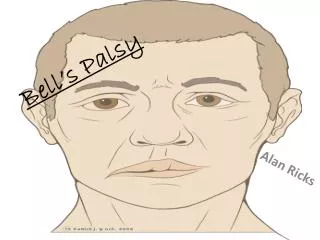
Bell’s Palsy. Department of Otorhinolaryngoglogy the 2nd Hospital affliatted to Medical college Zhejiang University Xu Yaping. Definition. Facial paralysis Acute onset, limited duration, minimal symptoms, spontaneous recovery Idiopathic in past Diagnosis of exclusion

E N D
Bell’s Palsy Department of Otorhinolaryngoglogy the 2nd Hospital affliatted to Medical college Zhejiang University Xu Yaping
Definition • Facial paralysis • Acute onset, limited duration, minimal symptoms, spontaneous recovery • Idiopathic in past • Diagnosis of exclusion • Most common diagnosis of acute facial paralysis
Etiology • Past theories: vascular vs. viral • McCormick (1972) – herpes simplex virus(HSV) • Murakami (1996) • 11/14 patients with HSV-1 in neural fluid • None in controls or Ramsay-Hunt syndrome • Temporal bone section at autopsy • Animal model inoculated with HSV-1
Natural History • Peiterson (1982): 1011 patients • Every decade of life, mean between 40-44 • 6-9% recurrent Bell’s palsy, M=W • Facial paresis (31%) -- 95% recover • Facial paralysis (69%) • 71% House-Brackmann grade I • 13% House-Brackmann grade II • 16% House-Brackmann grades III-V (fair-poor)
two prevailing theories • 1) vascular congestion with secondary ischemia to the nerve • 2) viral polycranioneuropathy. McGovernpostulated autonomic vascular instability with spasm of the nutrient arterioles. Thisvasospasmischemia nerve edemasecondary compression within the fallopian canal.
Evaluation • Careful history – timing • Associated symptoms (pain, dysgeusia) • SNHL, vesicles, severe pain • Trauma, acute or chronic OM, recurrent • Exposures • sudden oneset • absent of signs of central nervous system disease,ear disease, or cerebellopontine angle disease.
Physical exam: paralysis or paresis on one side of the face. House-brackmann Facial Nerve Grading System • normal: forehead,eye,mouth, • mild dysfunction • moderate dysfunction • moderately severe dysfunction • severe dysfunction • total paralysis
Audiometry • CT/MRI/other • Topographic • Electrophysiology
common complicated • exposure keratitis • inability to close the eyelid • diminished tearing • loss of corneal sensitivity
Anatomy • Intracranial • Meatal • Labyrinthine (2-4 mm) • Tympanic (11 mm) • Mastoid (13 mm) • Extracranial
Electrophysiology • Treatment plan based on 16% of patients who do not fully recover • Several tests used for prognosis • Measure amounts of neural degeneration occurred distal to injury by measuring muscle response to electrical stimulus • NET, MST, ENoG, EMG • Able to differentiate nerve fibers undergoing Wallerian degeneration
NET (nerve excitability test) Hilger first described in 1964 Compares current thresholds to elicit minimal muscle contraction 3.5 mA difference significant MST (maximum stimulation test) Compares responses generated with maximal electrical stimulation judged as difference in facial movement Absent or markedly decreased significant Electrophysiology
Electrophysiology • ENoG (electroneuronography) • Most accurate, objective • Records summation potential (CAP) • Degree of degeneration is directly proportional to amplitudes of measured potentials • Done after Wallerian degeneration starts (3-4 days) • Compare each day
Electrophysiology • ENoG • Esslen (1977) – over 90% degeneration on ENoG prognosis worsens • 90-97%: 30% recover fully • 98-99%: 14% recovery fully • 100%: none recovered fully • Fisch (1981) • 50% with 95-100% degeneration by 14 days have poor recovery • High likelihood of further degeneration if reaches 90% • Thus, if ENoG reaches 90% within 2 weeks: 50-50 recovery
Electrophysiology • EMG (electromyography) • Not useful in acute phase except as complementary test • Will be flat with neuropraxia, 100% degeneration, and early regeneration • Key in long-term evaluation (over 3 weeks) • Fibrillation potentials– degeneration • Polyphasic motor units– regenerating nerve
Medical Management • Eye protection • antimicrobial/ antiviral agents • Steroids: 7-10 days • Stankiewitz (1987)– no efficacy • Austin (1993)– randomized, double blind, placebo controlled study • Improvement in grade with prednisone • All with prednisone (House 1-2) • 17% without House 3 (statistically significant) • Trend towards denervation protection
Medical Management • Antivirals • Adour (1996)– double blind • Only 20% progressed to complete paralysis • Acyclovir had less degrees of facial weakness • Acyclovir had lower incidence of House 3-5 (House-Brackmann grade 3-5)
Surgical Management • Spirited debate over years • No surgery • Immediate decompression when total paralysis • Balance and Duel (1932)– first surgery • McNeill (1970)– no benefit (geniculate to stylomastoid foramen)– after 14 days
Surgical Management • Fisch and Esslen (1972)– 12 patients • Total facial nerve decompression via middle cranial fossa and transmastoid • Found conduction block at meatal foramen (94% patients) • Fisch (1981) • Decompression within 14 days for 90% degeneration for maximum benefit • May (1979) • Transmastoid decompression beneficial (decreased SF, Schirmer’s, MST reduced) • May (1984) • No patients benefited from surgery within 14 days
Prognosis • approximately 85% recover to normal within one year without treatment • The remaining 16% in this complete paralysis group have a fair to poor recovery • Approximately 6-9% develop recurrent Bell’s palsy. • Of those experiencing only a paresis, over 95% recover without sequelae
Hunt syndrome(Herpes zoster oticus) Definition • Herpes zoster is a viral disease most often affected sensory nerves due to involvement of theganglion. by Ramsay Hunt in 1910
intense ear pain and vesicle on EAC,concha • herpetic eruption over the drumhead,EAC and auricle. (ribs) • a facial nerve paralysis • vesicular eruptions on the head and neck • hearing loss and vertigo the trigeminal and audiotory nerves
treatment • narcotic analgesics for pain relief • oral steroids • antimicrobial/ antiviral agents • topical otic antibiotic-hydrocortisone solution • decompression when facial paralysis is beyond 60 days ( the horizontal segment and geniculate ganglion).
characters • recovery in days to weeks • the pain persist for several months