Download
1 / 43
551 likes | 1.26k Views
Physiology of The Small Intestine Motility and Secretion Dr. Mohammed Alzoghaibi. Learning Objectives . Motility in the small intestine. Control of intestinal motility. Secretions of the small intestine Digestion of carbohydrates, proteins and fats.

E N D
Physiology of The Small Intestine Motility and Secretion Dr. Mohammed Alzoghaibi
Learning Objectives • Motility in the small intestine. • Control of intestinal motility. • Secretions of the small intestine • Digestion of carbohydrates, proteins and fats. • Basic principles of gastrointestinal absorption. • Absorption of carbohydrates • Absorption of proteins • Absorption of fats • Absorption of fats • Absorption and secretion of electrolytes and water
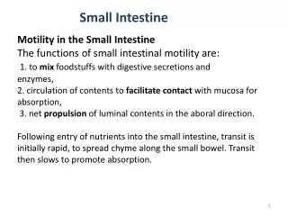
Motility in the Small Intestine • The movements of the small intestine can be divided into: • Segmenting (Mixing) contractions • Propulsive contractions (Peristalsis) • Migrating motor complex • Antiperistalsis • Peristaltic rush
Mixing (Segmentation) Contractions • When a portion of small intestine becomes distended, the segmentation contraction is activated by ENS to divide the intestine into spaced segments which last for fraction of min, and have the appearance of a chain of sausages. • As one set of segmentation contractions relaxes, a new set often begins at points between the previous ones. • The segmentation contractions become weak when the excitatory activity of ENS is blocked by the drug atropine. • The significance of segmentation contractions: • Blend different juices with the chyme • Bring products of digestion in contact with absorptive surfaces
Propulsive Movements (Peristalsis) • Propulsive movements can occur in any part of the small intestine, at a velocity of 0.5 to 2.0 cm/sec. • They are faster in the proximal intestine and slower in the terminal intestine. • They normally are very weak after traveling only 3 to 5 centimeters, and the net movement along the small intestine normally averages only 1 cm/min. This means that 3 to 5 hours are required for passage of chyme from the pylorus to the ileocecal valve.
Propulsive Movements (Cont.) • Organizes propulsion of material over variable distances within the intestinal lumen • Usual stimulus is distention • Myenteric plexus is important for these movements • It can be blocked by the drug atropine • Receiving segment ---contraction (longitudinal M.) ---relaxation (circular M.) • Propulsive segment ---contraction (circular M.) ----relaxation (longitudinal M.)
Migrating motor complex (MMC) • Bursts of depolarization accompanied by peristaltic contraction that begins in empty stomach during interdigestive period, travels a long whole length of small intestine to reach ileocaecal valve after 1.5-2 hour where it disappears. A new wave of MMC starts. • The activity of MMC terminates as soon as food is ingested. • The function of MMC is to propel any remnants in stomach & small intestine into colon during the interdigestive period.
Antiperistalsis Antiperistalsis contractionoccurs in the opposite direction between stomach and duodenum to allow more time for neutralization of chyme and between ileum and caecum to allow time for absorption. Peristaltic rush • Powerful rapid peristalsis due to intense irritation of intestinal mucosa as in infectious diarrhea. • Initiated mainly by extrinsic nervous reflexes to brain stem and back to gut. • Sweeps the contents of intestine into the colon and thereby relieving the small intestine of irritativechyme or excessive distension.
Movement of the villi The villous movement consists of fast shortening and slow lengthening as well as side to side movements. • Villous contractions are initiated by local nervous reflexes in response to chyme in small intestine. • They facilitate absorption and lymph flow from central lacteals into lymphatic system. • They are stimulated by villikinin hormone released by intestinal mucosa when it comes in contact with digestive products.
Control of intestinal motility Neural factors: • Vagal excitation increases intestinal and villous movements. • Sympathetic excitation decreases intestinal and villous movements. • Gastroileal reflex is initiated by gastric distension. Impulses are conducted through myenteric plexus to initiate a fast peristaltic wave passing to the ileum. The ileocaecal valve relaxes allowing chyme to pass into cecum. This reflex is mediated by vagus nerve.
Control of intestinal motility (cont.) Hormonal factors • Gastrin, CCK, insulin and serotonin stimulate intestinal motility. Gastrin and CCK relax ileocaecal sphincter. • Motilin secreted from duodenum stimulates intestinal motility and regulate MMC. • Secretin and glucagons inhibits intestinal motility and contract ileocaecal sphincter. • Villikinin stimulates movement of the villi.
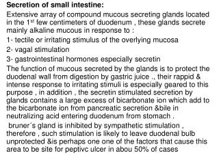
Secretions of The Small Intestine Secretion of Mucus by Brunner’s Glands in the Duodenum • Brunner’s glands are located in the wall of the first few centimeters of the duodenum. They secrete large amounts of alkaline mucus, which contains a large amount of bicarbonate ions, in response to (1) irritating stimuli on the duodenal mucosa; (2) vagal stimulation, (3) secretin. • Mucus protects the mucosa. • Brunner’s glands are inhibited by sympathetic stimulation
Secretion of Intestinal Juices by the Crypts of Lieberkühn • Crypts of Lieberkühnare small pits which lie between intestinal villi. • The surfaces of both the crypts and the villi are covered by an epithelium composed of 2 types of cells: (1) goblet cells, secrete mucus, and (2) enterocytes, secrete large quantities of H2O and electrolytes and over the surfaces of adjacent villi, reabsorb H2O, electrolytes & end products of digestion.
Digestive Enzymes in the Small Intestinal Secretion • Most of the enzymes are found either in the brush border or in the cytoplasm of the enterocytes. The enzymes that are secreted into the lumen are enteropeptidase and amylase. • The enterocytes of the mucosa contain digestive enzymes. These enzymes are the following: (1) Aminopeptidases, Oligopeptidases, Intracellular di- and tripeptidases for splitting small peptides into amino acids. (2) Four enzymes: sucrase, maltase, isomaltase, and lactase—for splitting disaccharides into monosaccharides. (3) Small amounts of intestinal lipase for splitting neutral fats into glycerol and fatty acids. (4) Nucleotidases for splitting nucleotides into purine and pyrimidine bases, phosphoric acid and pentose sugar.
Control of intestinal secretion • Brunner's gland secretion is stimulated by secretin, tactile and vagal stimulation. • Intestinal juice secretion is stimulated by: • a. Distension, tactile and irritating stimuli. • b. Hormones as gastrin, secretin, CCK, glucagons, enterocrinin. • c. Sympathetic stimulation exerts an inhibitory effect.
Digestion of Carbohydrates in the Mouth and Stomach • The ptyalin (an α-amylase) enzyme in saliva hydrolyzes starch into the disaccharide maltose and other small polymers of glucose. • The starch digestion sometimes continues in the body and fundus of the stomach for as long as 1 hour before the food becomes mixed with the stomach secretions.
Digestion of Carbohydrates in the Small Intestine Digestion by Pancreatic Amylase. • Pancreatic secretion has α-amylase that is almost identical in its function with the α-amylase of saliva but more powerful. Therefore, within 15 to 30 minutes after the chyme empties from the stomach into the duodenum and mixes with pancreatic juice, virtually all the carbohydrates will have become digested. • The carbohydrates are almost totally converted into maltose and/or other very small glucose polymers before passing beyond the duodenum or upper jejunum.
Hydrolysis of Disaccharides by Intestinal Enzymes • The enterocytes lining the villi of the small intestine contain four enzymes (lactase, sucrase, maltase, and α-dextrinase), which are capable of splitting the disaccharides lactose, sucrose, and maltose, plus other small glucose polymers, into their constituent monosaccharides. • These enzymes are located in the enterocytes covering the intestinal microvilli brush border, so that the disaccharides are digested as they come in contact with these enterocytes.
Digestion of Proteins in the Stomach • Pepsin is the important peptic enzyme of the stomach (active at a pH=2.0 - 3.0 and is inactive at a pH above about 5.0). • The pH of the stomach averages around 2.0 - 3.0. • One of the important features of pepsin digestion is its ability to digest the protein collagen. • Collagen is a major constituent of the intercellular connective tissue of meats; therefore, for the digestive enzymes of the digestive tract to penetrate meats and digest the other meat proteins, it is first necessary that the collagen fibers be digested. • Pepsin only initiates the process of protein digestion, usually providing only 10 to 20 per cent of the total protein digestion.
Digestion of Proteins by Pancreatic Secretions • Most protein digestion occurs in the duodenum and jejunum. • Both trypsin and chymotrypsin split protein molecules into small polypeptides; carboxypolypeptidase then cleaves individual AA from the carboxyl ends of the polypeptides. • Proelastase is converted into elastase, which then digests elastin fibers that partially hold meats together. • Only a small percentage of the proteins are digested all the way to their constituent AA by the pancreatic juices. • Most remain as dipeptides and tripeptides to be digested by Peptidases in the Enterocytes mainly in the duodenum and jejunum.
Digestion of Fats in the Intestine • Less than 10 % of triglycerides is digested in the stomach by lingual lipase. • All fat digestion occurs in the small intestine. Emulsification of Fat by Bile Acids • Break the fat globules into very small sizes under the influence of bile salts , so that the water-soluble digestive enzymes can act on the globule surfaces (emulsification of the fat). • The polar parts (the points where ionization occurs in water) of the bile salts and lecithin molecules are highly soluble in water. So the they are amphipathic molecules. • The major function of the bile salts and lecithin, especially the lecithin, in the bile is to make the fat globules readily fragmentable by agitation with the water in the small bowel.
Role of Bile Salts to Accelerate Fat Digestion Formation of Micelles • Bile salts have the ability to form micelles, (each bile salt molecule is composed of a sterol nucleus that is fat-soluble and a polar group that is water-soluble • Micelles are small spherical, cylindrical globules 3 to 6 nm in diameter composed of 20 to 40 molecules of bile salt. • The polar groups are (-) charged, they allow the entire micelle globule to dissolve in the water of the digestive fluids and to remain in stable solution. • The micelles act as a transport medium to carry the monoglycerides and free fatty acids to the brush borders of the intestinal epithelial cells.
Digestion of Triglycerides by Pancreatic Lipase • The most important enzyme for digestion of the triglycerides is pancreatic lipase. End Products of Fat Digestion
Absorptive Surface of the Small Intestinal Mucosa Villi • The absorptive surface of the small intestinal mucosa, showing many folds called valvulaeconniventes (villi). • They increase the surface area of the absorptive mucosa about threefold. • They are well developed in the duodenum and jejunum. • The presence of villi on the mucosal surface enhances the total absorptive area another 10-fold. Longitudinal section of the small intestine, showing the valvulaeconniventes covered by villi
Absorptive Surface of the Small Intestinal Mucosa Villi • The epithelial cell on each villus is characterized by a brush border, consisting of as many as 1000 microvilli protruding into the intestinal chyme (increases the surface area another 20-fold). Brush border of a gastrointestinal epithelial cell
Absorption of Carbohydrates • All the carbohydrates in the food are absorbed in the form of monosaccharides; only a small fraction are absorbed as disaccharides. • Glucose and galactose absorption occurs in a cotransport mode with active transport of Na+ (2ry active transport) . • Fructose is independent on Na+ but it transports in lumenal membrane via facilitated diffusion. • Pentose is transported by passive diffusion
Proteins are absorbed in the form of dipeptides, tripeptides, and a few free amino acids. • L- AA are transported by 2ry active transport. • Di and tripeptides cross the brush border by active transport protein carrier. They are hydrolyzed by brush border and cytoplasmic oligopeptidases. • AA leaves the cell at the basolateral membrane by facilitated transport. Absorption of Proteins
Absorption of Fats In the presence of an abundance of bile micelles, about 97 per cent of the fat is absorbed; in the absence of the bile micelles, only 40 to 50 per cent can be absorbed.
Absorption of vitamins • Fat-soluble vitamins (A, D, E, & K) are incorporated into micelles and absorbed along with other lipids • Most water-soluble vitamins (C, B1, B2, B6, and folic acid) are absorbed by Na-dependent co-transport mechanisms • Vitamin B12 is absorbed in the ileum and requires intrinsic factor • Gastrectomy results in the loss of parietal cells and loss of intrinsic factor pernicious anemia
Absorption and secretion of electrolytes and water • Electrolytes and H2O may cross intestinal epithelial cells by either cellular or paracellular • The permeability of the tight junctions varies with the type of epithelium • A tight epithelium is in the colon • Leaky epithelia are the small intestine and gallbladder
Absorption of Na+ Na moves into the intestinal cells by the following mechanisms: • Passive diffusion • Na-glucose or Na-amino acid co-transport • Na-Cl exchange • Na-H exchange • The next step in the transport process is osmosis of water into the paracellular spaces because a large osmotic gradient has been created by the elevated concentration of ions in the paracellular space. • Aldosterone Greatly Enhances Sodium Absorption: This effect of aldosterone is especially important in the colon because it allows virtually no loss of NaCl and water.
Absorption of Cl- • Cl- absorption accompanies Na+ absorption by the following mechanisms: • Passive diffusion • Na-Clcotransport • Cl-HCO3 exchange Absorption and secretion of K • K is absorbed in the small intestine by passive diffusion • K secretion in the colon is stimulated by aldosterone • Excessive loss of k in diarrheal fluids causes hypokalemia
Secretion of Bicarbonate Ions in the Ileum • The epithelial cells on the surfaces of the villi in the ileum and large intestine have a special capability of secreting bicarbonate ions in exchange for absorption of chloride ions. This is important because it provides alkaline bicarbonate ions that neutralize acid products formed by bacteria in the large intestine.
Ca++Absorption by Enterocytes • plasma Ca parathyroid hormone 25-hydroxy-vitamin D3kidney 1,25 dihydroxy-vitamin D3 Stimulates synthesis of Ca-binding protein and Ca-ATPase in enterocytes
Hormonal control of absorption & secretion • Glucocorticoid = absorption of H2O & ions (small & large intestine) • Catecholamines = intestinal secretion • Somatostatin = H2O & ions absorption (ileum & colon) • Epinephrine = NaCl absorption (ileum) • Aldosterone = synthesis of Na channel (colon)