Download
1 / 55
670 likes | 1.5k Views
Common Foot Problems. Garrett Post, MD PGY-2 Emory Family Medicine Residency Program March 20, 2008. Common Foot Problems. Anatomy – Bones. Anatomy - Bones. Anatomy - Bones. Anatomy - Vascular. Anatomy - Nervous. Anatomy - Nervous. Anatomy - Musculoskelatal.

E N D
Common Foot Problems Garrett Post, MD PGY-2 Emory Family Medicine Residency Program March 20, 2008
Common Foot Problems Anatomy – Bones
Anatomy - Musculoskelatal Where does the Achilles tendon derives its name? Hint:
Greek Mythology Thetis tried to immortalize Achilles by dipping him into the sacred river Styx. What sign is indicative of an Achilles tendon rupture?
Greek Mythology Thetis tried to immortalize Achilles by dipping him into the sacred river Styx. What sign is indicative of an Achilles tendon rupture? Thompson’s sign
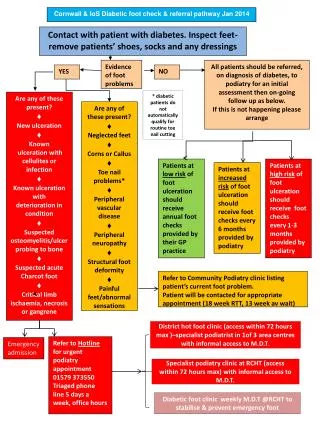
What is wrong with my Foot?!? • Fungal and Bacterial Conditions • Corns and calluses • Warts • Bunions • Ingrown toenails • Hammer toe • Spurs • Plantar Fasciitis • Ankle Sprain • Morton’s Neuroma • Post-tib Tendinitis • Sesamoiditis • Shin Splints • Tarsal Tunnel Syndrome • Metatarsalgia • Excessive Pronation • Excessive Supination • Achilles tendinitis • Gout
What is wrong with my Foot?!? • Fungal and Bacterial Conditions • Corns and calluses • Warts • Bunions • Metatarsalgia • Hammer toe • Spurs • Plantar Fasciitis • Ankle Sprain • Morton’s Neuroma • Post-tib Tendinitis • Sesamoiditis • Shin Splints • Tarsal Tunnel Syndrome • Ingrown toenails • Excessive Pronation • Excessive Supination • Achilles tendinitis • Gout
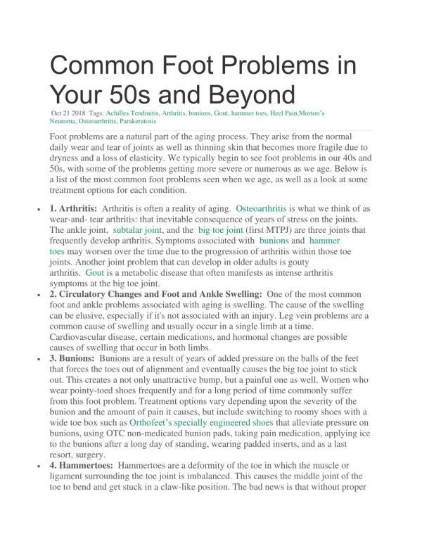
Bunions - Hallux Valgus Not so much “good luck”… But you can cross them anyway.
Bunions - Hallux Valgus • NOT likely from high heel shoes (Myth) • High heels may exacerbate the underlying etiology.
Hallux Valgus • Affects 1% of adults in the US. (National Health Interview survey by the National Center for Health Statistics) • Incidence increased with age • 3% 15-30 years • 9% 31-60 years • 16% > 60 years • Female to Male ratio - 2:1 to 4:1 • The role of genetic predisposition has also been noted, with evidence to suggest familial tendencies. Gould et al., 1980 1
Hallux Valgus - Etiology Biomechanical instability • Most common • Difficult to understand and evaluate • Genetically inherited and/or from underlying conditions
Hallux Valgus - Etiology • Arthritic/metabolic conditions • Gouty arthritis • Rheumatoid arthritis • Psoriatic arthritis • Connective tissue disorders such as Ehlers-Danlos syndrome, Marfan syndrome, Down syndrome, and ligamentous laxity
Hallux Valgus - Etiology • Neuromuscular disease • Multiple sclerosis • Charcot-Marie-Tooth disease • Cerebral palsy • Traumatic compromise • Structural deformity i.e. Abnormal metatarsal length
Bunions - Pathophysiology • Hallux and digits generally remain parallel to the long axis of the foot. • Medial tension causes the medial collateral ligaments to pull on the dorsomedial aspect of the first metatarsal head, causing the bone to proliferate. • Lateral tension causes the sesamoid apparatus to fixate in a laterally dislocated position. • Remodeling (bone and cartilage) occurs laterally and medially. • Without correction of the biomechanical etiology, excessive pronation continues, with propagation of the deformity.
Presentation • Hallux position • Medial prominence • First metatarsophalangeal joint ROM • First ray ROM • Plantar keratosis • Pain location • Contracture of the extensor hallucis longus • Associated deformities
Evaluation – Labs and XRay • Uric acid, ESR, CRP, ANA, RF • Foot AP, Lat, and Oblique
Staging • Stage 1 - excessive pronation causes hypermobility of the first ray, causing the tibial sesamoid ligament to be stretched and the fibular sesamoid ligament to contract, and lateral subluxation of the proximal phalanx occurs. • Stage 2 - hallux abduction progresses, with the flexor hallucis longus and flexor hallucis brevis gaining lateral mechanical advantage. • Stage 3 - further subluxation occurs at the first metatarsophalangeal joint, with formation of metatarsus primus adductus. • Stage 4 - the first metatarsophalangeal joint finally dislocates.
Hallux Valgus - Treatment • Medical therapy • Adapting footwear • Pharmacologic or physical therapy • Functional orthotic therapy • Activity • Weight • Top cover • Biomechanical examination • Modifications - cushions • Surgical therapy
Recovery • Six months to full activity • Best to take 2 weeks off and elevate leg • Ice popliteal fossa for 1 week postoperatively to reduce swelling • Ambulating in 2 weeks • Pain free without strenuous exercise at 3-5 weeks
Plantar Fasciitis In US5: • 10% of runner-related injuries • 11-15% of all foot symptoms requiring professional care • 10% - general population • Presents bilaterally – 1/3 cases • Race and ethnicity play no role in the incidence • Peak incidence may occur in women aged 40-60 years.
Plantar Fasciitis • A thickened fibrous aponeurosis • Originates from medial tubercle of the calcaneus • Runs forward to insert into the deep short transverse ligaments of the metatarsal heads • Continues forward to form the fibrous flexor sheathes on the plantar aspect of the toes • Central plantar fascia is the thickest and strongest section, and most likely to be involved with plantar fasciitis. • Function - provide static support for the longitudinal arch of the foot and to assist with shock absorption during foot strike (2-3 times an individual's body weight with each step).
Plantar Fasciitis - Pathophysiology • Increased risk – • pes planus (low arches or flat feet) • pes cavus (high arches) • Pain - collagen degeneration associated with repetitive microtears of the plantar fascia. • Common h/o increase in weight-bearing activities causing the microtears exceeding healing capacity. • Elderly – biomechanical (functional and degenerative factors)
Plantar Fasciitis - Etiology Extrinsic risk factors • Training errors • Overuse – Most common • Equipment Intrinsic risk factors • Structural risk factors • Overpronation, discrepancy in leg length, excessive lateral tibial torsion and excessive femoral anteversion • Functional risk factors • Tightness or weakness of soleus, gastrocnemius, Achilles tendon and intrinsic foot muscles. • Degenerative risk factors • Poor intrinsic muscle strength and poor force attenuation secondary to acquired flat feet and compounded by a decrease in the body's healing capacity
Plantar Fasciitis - Symptoms Intense sharp heel pain with the first couple of steps in the morning. Pain is anterior aspect of the calcaneus, but may radiate proximally in more severe cases. Dull ache in the heel at the end of the day, especially after extensive walking or standing.
Plantar Fasciitis - Physical Exam • Point of maximal tenderness at the anteromedial region of the calcaneus. • Pain along the proximal plantar fascia.
Plantar Fasciitis - Physical Exam • "windlass" test - Passive dorsiflexion of the toes which elicits Sx pain • Having the patient bear weight during the windlass test increased the sensitivity of the test from 13.6% to 31.8% (De Garceau, 2003) 9
Plantar Fasciitis - Treatment • Correcting training errors by: • relative rest • ice after activities • evaluation of the patient's shoes and activities • Correction of biomechanical factors by: • stretching and strengthening program (PT) • Then, consider night splints and orthotics • Finally, other treatment options are considered. NSAIDs are considered throughout the treatment course for pain control. Time for resolution - often 6 to 18 months.
Plantar Fasciitis - Treatment Iontophoresis – Electric stimulation • Use of electric impulses from a low-voltage galvanic current stimulation unit to drive topical corticosteroids into soft tissue structures. Corticosteroid Injections • Greatest benefit if administered early • Studies7,8 have found steroid treatments successful 70% or better • Potential risks • rupture of the plantar fascia • fat pad atrophy. Autologous blood injections • Ignites the healing process Surgery • Success rate of surgical release is 70% to 90% • Potential risks • flattening of the longitudinal arch • heel hypoesthesia.
Ankle Sprain Common site for acute musculoskeletal injuries. • Sprains – 75% of ankle injuries • Acute ankle trauma – 10% to 30% of sports-related injuries in young athletes • Estimated 1 million persons present with acute ankle injuries • >40% ankle sprains have potential to cause chronic problems
Ankle Sprain • Anterior drawer test - assess the integrity of the anterior talofibular ligament • Inversion stress test - assess the integrity of the calcaneofibular ligament
Ankle Sprain GradeI: partial tear of a ligament • Mild tenderness and swelling • Slight or no functional loss (i.e., patient is able to bear weight and ambulate with minimal pain) • No mechanical instability (negative clinical stress examination) Grade II: incomplete tear of a ligament, with moderate functional impairment • Moderate pain and swelling • Mild to moderate ecchymosis • Tenderness over involved structures • Some loss of motion and function (i.e., patient has pain with weight-bearing and ambulation) • Mild to moderate instability (mild unilateral positivity of clinical stress examination) Grade III: complete tear and loss of integrity of a ligament • Severe swelling (more than 4 cm about the fibula) • Severe ecchymosis • Loss of function and motion (i.e., patient is unable to bear weight or ambulate) • Mechanical instability (moderate to severe positivity of clinical stress examination)
Ankle Sprain Grades I or II sprains • Accurate early diagnosis • RICE (rest, ice, compression and elevation) • Maintenance of ROM - Functional Rehabilitation • Ankle support Grade III • Early motion and mobility are recommended • Ligamentous strength returns months after injury – delayed functional rehab • May require surgical intervention
Ottawa ankle and foot rules • XR ankle indicated if • pain in the malleolar zone and • any of these findings: • bone tenderness at A, • bone tenderness at B or • inability to bear weight immediately and in the emergency department. • pain in the midfoot zone and • any of these findings: • bone tenderness at C, • bone tenderness at D or • inability to bear weight immediately and in the emergency department. Sensitivity 100% for malleolar fractures (95 percent confidence interval [CI]; range: 82 to 100%) Sensitivity 100% for midfoot fractures (95 percent CI; range: 95 to 100%) 6
Ankle Sprain • Sx more than six weeks – CT or MRI to rule out talar dome lesions
Ingrown toenails Etiology4 • Inheritance - genetically predisposed inwardly curved nails with distortion of one or both nail margins • Underlying bony pathology causing deformation of the nail • Obesity “fat feet” - deepening of the nail groove • HIV antiviral therapy - increased incidence of ingrown nails • Prior trauma - irregularly shaped nail
Ingrown toenails Stage 1 – • erythema, • slight edema, and • pain with pressure to the lateral nail fold.