Download

1 / 102
1.03k likes | 1.24k Views
Eye Lids. خروج. السلام عليكم ورحمة الله وبركاته اولا دعوني ارحب بكم فأهلا بكم في برنامجي البسيط التي اتمنى من الله عز وجل ان اكون قد وفقت في تقديمه باسهل الطرق..

E N D
خروج السلام عليكم ورحمة الله وبركاته اولا دعوني ارحب بكم فأهلا بكم في برنامجي البسيط التي اتمنى من الله عز وجل ان اكون قد وفقت في تقديمه باسهل الطرق.. وأحب ان اعلم الجميع اني وضعت خلف بعض المصطلحات صور .. فبمجرد الضغط على اللون الأزرق كن يقينا ان البرنامج سوف يحولك لصفحة تحتوي على الصور لتسهيل عليك تخيل منظر المرض..وللرجوع للصفحة السابقة اضغط على سهم الرجوع ... وان اتركك لتشاهد البرنامج مع تمنياتي لكم بالتوفيق الرجاء قراءة الملاحظات قبل مشاهدة البرنامج
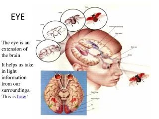
خروج TEXT BOOK OF DISEASES OF THE EYE The eye lid is made up of 4 layers. They are from without in-wards: 1) Skin – it is thin and characterized by absence of fat. 2) Muscle layer. a) Orbicular is oculi consists of horizontal concentric fibers it is supplied by the zygomatic branch of the facial nerve. When the orbicularis oculi contracts the lids are firmly closed. b) Levator palpebrae superiors. The muscle fibres are arranged vertically; they end in an aponeurosis which is inserted . i) To the skin of the upper lid. ii) Upper border of the tarsus. iii) Conjunctiva at the fornix. It is supplied by the upper division of the 3rd cranial nerve. It raises the upper lid. c) Muller's muscle: supplied by the sympathetic nerve. 3) Tarsus : Consists of dense fibrous tissue. Embedded in it are enormously developed sebaceous glands-the meibomian (tarsal) glands. 4) Mucous layer : formed by the palpebra conjunctiva.
خروج Glands of the eye lids: • Meibomian (tarsal) glands : They are embedded in the tarsus and are modified sebaceous glands. They secrete an oily secretion. They open through vertically arranged ducts into the lid margin. • Glands of Zeis : they are sebaceous glands developed as outgrowth of the hair follicles of the eye lashes. They are situated at the lid margin. • Glands of Moll: these are modified sweat glands. The lid margin – is covered with stratified epithelium which forms a transition between the skin and the conjunctiva. It consists of: • Eye lashes – arranged in 2 – 3 rows anteriorly. • Opening of the ducts of the meibomian gland posteriorly. • Glands of Zeis and Moll.
خروج • DISEASES OF THE LIDS: Oedema of lids – this is common and owing to looseness of the tissue may be so great as to close the eye. • ANATOMY AND DISEASES OF THE LIDS Causes: • Inflammatory oedema. • Inflammation of the lids – allergic dermatitis due to atropine ointment and cosmetics, stye, insect bite. • Acute conjunctivitis. • Acute dacryocystitis. • Acute iridocyclitis. • Panophthalmitis. • Orbital cellulitis. • Passive oederma due to circulatory obstruction. • Nephritic syndrome. • Cardiac failure. • Cavernous sinus thrombosis.
خروج • INFLAMMATION OF THE LIDS:- Blepharitis: It is a chronic inflammation of the margin of the lid. The lid margin becomes thickened and red. • VARIETIES AND ETIOLOGY: 1) Squamous blepharitis: there is encrustration of lid margin by white scales. It is of 2 types. • Oleosa – Or seborrheic : Often essentially metabolic associated frequently with seborrheic dermatitis of scalp (dandruff). Exacerbation and remission of the ocular lesions parallel those of lesions of the scalp. b) Siccaa : Chemical irritants and cosmetics such as "surma", acne rosacea, uncorrected refractive error particularly astigmatism.
خروج 2) Ulcerative blepharitis :There is encrustation of the lid margin by yellow scales with underlying ulceration. The ulcers bleed on removing the scales. This distinguishes the condition from matting together of the lashes by conjunctival discharge in conjunctivitis; removal of the crusts in conjunctivitis reveals normal lid margin. Ulcerative blepharitis is an infective condition caused by staphylococci. 3) Mixed blepharitis:Staphylococci infection super imposed on Squamous blepharitis. 4) Margin is associated: with angular conjunctivitis – foamy discharge and excoriation of the skin of the lateral and medial canthi. It is caused by Morax – Axenfeld bacillius.
خروج • TEXT BOOK OFDISEASES OF THE EYE The patients are usually children debilitated from : 1) Living under poor hygienic conditions – exposure to dust, smoke. 2) DISEASES – exanthematous disease, upper respiratory infections, tuberculosis, diabetes. 3) Dietary deficiency – malnutrition. Occasionally parasite causes blepharitis, blepharitis acarica. • Symptoms : Itching, soreness, lacrimation, photophobia.
خروج • Sequelae or complications: • Chronic conjunctivitis. • Marginal corneal ulcer. • Styes. • Permanent loss of a greater or losser number of lashes (madarosis ) due to destraction of root of the cilia. • Tylosis – usuall affects upper lid. There is hypertrophy of lid border causin this part to become rounded and thick and to droop on account of its own weight. • Trichiasis. • Entropion. • Ectropion.
خروج • Treatment : • Improvement of general health and living condition. • Treatment of seborrheic dermatitis o scalp with shampoo. • Correction of refractive error. • Removal of scales and crust with warm 3% soda bicarb solution. • In cases of squamous blepharitis, dilute baby shampoo is applied on the lid margin with a swab stik. In more obstinate lesions, ointment of selenium oxide is used. • allergy to chemical and cosmetic, 1% hydrocortisone ointment is applied to the lid margin thrice a day. • In cases of ulcerative blepharitis antibiotics such as chloromycetin, erythoromycin or tetracycline are applied to the lid margin. In more severe cases the antibiotic which has proved to be effective is given systemically, in addition to local treatment. • In cases of angular blepharitisoxteracycline ointment is applied.
خروج • INFLAMMATION OF THE GLANDS OF THE LIDS : 1) Eternal hordeolumor stye : is a circumscribed, acute inflammation at the edge of the lid, caused by staphylococcal infection of the glands of Zeis usually ending in suppuration. • Etiology : Most common in children and young adults, often appear in crops. Frequently associated with blepharitis or lowered state of health – diabetes mellitus, and uncorrected refractive error. • Symptorns and Signs: Red swelling appears in the lash line of the margin of the lid, accompanied by pain, tenderness and often by considerable oedema of the lids. Very soon a yellowish summit will be seen orderlid indicating suppuration.
خروج • Treatment : • Hot fomentation to hasten suppuration : as soon as a yellow spot is seen, the pus should be evacuated by a horizontal incision at the lid margin or epilation. • Antibiotics – choromycetin ointment. • Analgesics. • Prophylaxis. • Antibiotic ointment. • Treatment of diabetes, blepharitis, correction of refractive errors. • Avoidance of excess of sweets, oil in the diet.
خروج 2) Hordeolum internum: this is an acute suppurative inflammation of a meibomian gland due to staphylococcus. Sometimes it may be due to secondary infection of a chalazion. - Symptoms and Signs: Symptoms are more violent than those of stye. Very soon pus points on the palpebral conjunctiva. • ANATOMY AND DISEASES OF THE LIDS 33 Treatment : • It may be removed by operation – incision and curettage through the conjunctiva. A vertical incision is made through the palpebral conjunctiva and with a chalazion curette the contents are removed and the walls thoroughly scraped. Vertical incision is made to avoid cutting ducts of neighbouting meibomian glands and to prevent Entropion. • Injection of cortisone into the chalazion can be used as an alternate fromof treatment.
خروج • Blepharospasm : It is a condition in which there are involuntary and forcible eyelid closure (Refer Corneal ulcer for difference from photophobia). 1) Reflex sensory stimulation through branches of 5th cranial nerve – commonest. a) Phlyctenular keratoconjunctivitis. b) Foreign body on cornea. c) Membranous and pseudo membranous – conjunctivitis. d) Acte iridocyclitis. 2) Excessive stimulation of retina. a) Bright light on sensitive eye. b) Dilated pupil. c) Albinism. 3) Essential Blepharospasmwithout any cause. 4) Hysteria.
خروج • Trichiasis : - is an inversion of a varying number of eye lashes so that they rub against the conjunctiva or cornea. The margin of the lid may have a normal position, the displacement affecting only the lashes or the margins may be turned inward (Entropion ) as well as the lashes. • Etiology : same as Entropion. • Signs and symptoms : (same for Entropion). The misdirected lashes cause mechanical irritation and injury to cornea with ulceration – pain, lacrimation, photophobia, Blepharospasm, vascularisation and opacities of cornea. • Treatment : If only a few cilia involved – epilation or electrolysis to destroy the hair follicles. If extensive – surgical treatment as for cicatricial Entropion.
خروج • TEXT BOOK OF DISEASES OF THE EYE • Treatment : -is the same as same as for stye except that the incision should be made exactly as for chalazion i.e. a vertical incision on the palpebral conjunctiva. This is to avoid cutting ducts of neighbouring meibomian glands and to prevent Entropion. • chalazion : -This is a chronic granulomatous enlargement of one of the meibomain glands. • Etiology & Pathology : It occurs most frequently in adults. The meibomian duct becomes obstructed through proliferation of its epithelium and consequently the gland enlarges. The fatty secretion escapes into the surrounding tissue and excites a foreign body reaction (lipogranuloma) which consists of lymphocytes, epitheloid cells and giant cells. The blood supply is cut off by the surrounding fibrous tissue leading to degeneration of contents – jelly like mass.
خروج • Symptoms : chalazionmay cause: 1) Cosmetic disfigurement – lid swelling. 2) Conjunctiva irrition. • Signs : The process develops slowly. After weeks or months it presents a circumscribed swelling which feels hard, not adherent to skin. On everting the lid, its situation is usually shown by a purple discolouration of the conjunctiva.
خروج • Complications: • It may be secondarily infected forming an internal hordeolum. • In old people, recurrent chalazion may lead to the development of Meibomian gland carcinoma. • Mechanical ptosis in cases of large chalazion. • The chalazion may press on the cornea causing astigmatism. • The chalazion may burst either on the skin surface or on the conjunctival surface, with granulation tissue protruding. • The granuloma may protrude through a duct of the meibomian gland on the lid margin – marginal chalazion.
خروج • Entropion: is a rolling of the margin of the lid and with it the lashes. Etiology and Varieties : I) Cicatricial entopion – due to cicatricial changes in the conjunctiva and distortion of the tarsal plate, most commonly affects the upper lid . a) Old case of trachoma . b) Blepharitis c) Burns and other injuies to the lids d) Operation upon the lids e) Diphtheritic conjunetivitis
خروج II) Spastic entropion: Due to spasm of the palpebral portion of the orbicularis muscle, most commonly affecting lower lid, strong contraction of the circularly arranged fibers tends not only to approximate the lid margins but also to turn them inwards of outwards according to the degree of mechanical support afforded by the globe and orbital contents. If the support is insufficient intropion is produced: i) Atrophy or absence of eye ball ii) Old persons ( senile entropion) owing to absence of orbital fat. iii) Tight bandaging after surgical operation of the eye. iv) Blepharospasm Symptoms and signs – same as for trichiasis .
خروج Treatment: I) Cicatricial entrpion : The principles governing various operation are: i) Altering the direction of eyelashes a) Snellen,s operation b) Jaesch Arlt operation ii) Straightening of the distorted tarsus. II) Spastic entropion i) Pull the lower lid down and out and apply leucoplast which should extend below the mandible. ii) Injection of alcohol is made close to the lateral 3rd of the lid margin . • Plastic surgery of lower lid - modified wheeler's operation Ectropion : It is a rolling out of the margin of the lid Etiology and varieties : Cicartical Ectropion : due to cicatricial changes in the skin of the lids- Blepharitis, burns, operations on the lids, leprosy. Both upper and lower lids may be affected.
خروج 2) Senile ectropion – the lower lid is affected in old age due to laxity of the tissue of the lid and due to loss of tone of the orbicularis muscle. 3) Paralytic ectropion – it occurs as a result of weakness of the orbicularis muscle due to facial nerve paralysis . The lower lid is affected. Symptoms – Epiphora due to eversion of lacrimal punta. Complications 1) Xerosis of conjunctiva 2) Chronic conjunctivitis and exposure keratitis particularly in Ectropion of upper lid. Treatment : 1) In mild cases row of heat cautery applied to the palpebral conjunctiva below the lek margin . As a result of fibrosis of conjunctiva ectropion is corrected . 2) Lateral tarsorrhaphy 3) In severe case, platstic surgery – Kuhnt Szymanowski operation.
خروج SYMBLEPHARON: is a cicatricial attachment between the conjunctiva of the lid and the eye ball, it may affect both lids, but usually the lower, sometimes it includes part of the cornea. Tyes: 1) Anterior- when extending bridge like from lid to globe, leaving a free portion of the conjunctiva corresponding to the fornix . 2) Posterior- When it involves only the fornix. 3) Total – When the lids are adherent to the globe throughout. Etiology : It is caused by the junction of 2 opposing granulating surfaces raw surfaces hence it occurs after. 1) injuries especially burns from lime, acids, and molten metal. 2) Operations. 3) Trachoma, rarely diphtheritic conjunctigitis.
خروج Symptoms: 1) It often interferes with movement of eye ball producing diplopia. 2) Traction upon the adherent parts causes irrition 3) In extensive cases the cornea is involved and vision is affected. 4) If there is inability to close , lagoph thalmos. 5) Cosmetic disfigurement. Treatment: 1) Prophylactic - use of contact shell in fresh cases of alkali burns, after operations on lids. 2) Curative. a) If anterior and not extensive, the band is divided and the 2 raw surfaces kept from uniting by separating them daily with a glass rod smeared with antibiotic ointment . b) In more sever cases of anterior symblepharon, in posterior and total symblepharon the separated raw surfaces must be cobered with conjunctiva or with grafts of mucous membrane from the lip to keep them from uniting .
خروج Lagophthalmos: This is the condition of incomplete closure of the palpebral aperture when an attempt is made to shut the eyes. Causes: 1) Exophthalomos as in Graves' disease, proptosis due to orbital tumour. 2) Facial never paralysis . 3) Cicatricial ectropion of upper lid . 4) Symblepharon. 5) Laxity of the tissue and absence of reflex blinking in people who are extremely ill – coma, keratomalacia etc. Complications: 1) Parenchymatous xerosis of conjunctiva. 2) Chronic conjunctivitis and exposure keratitis Treatment: 1) Of the cause whenever possible. 2) Application of a bland or antibiotic ointment to protect cornea. 3) Lateral or median tarsorrhaphy to protect the cornea.
خروج 1) Distichiasis: It is a condition in which there is an extra posterior row of eye lashes. The posterior row occupies the position of the opening of Meibomian glands, these lashes may irritate the cornea. 2) Coloboma of the lid: It is a triangular gap in the lid magin, generally affecting the upper lid. It may form part of Golden Har's syndrome- accessory auricles, dermolipoma at the limbus, hypoplasia of the maxilla and anomalies of the vertebral column. • ptosis or blepharoptosis : is a drooping of the upper lid usually due to defective development or paralysis of the levator palpebrae superioris (L.P.S.). All degrees o ptosis occur- partial or complete. When severe it interferes with vision by covering the pupil. 1) Patients attempt to raise the lid by forced action of the occipito frontalis muscle, wrinkling the skin of the forehead and raising the brow. 2) When condition is severe and bilateral they favour exposure of the pupil by throwing the head backwards.
خروج Etiology & Classification: 1) Congenital ptosis: a) With normal superior rectus funtction. b) With superior rectus weakness- developmentally superior rectus is closely related to L.P.S. • Marcus Gunn or jaw-winking ptosis. 2) Acquired ptosis: • Neurogenic – due to lesion of 3rd never nucleus, 3rd nerve trunk in its intracranial or orbital course as in cases of brain tumour, meningitis, aneurysm etc. - Horner's syndrome due to affection of sympathetic nerves. b) Myogenic – Myasthenia gravis. c) Mechanical ptosis - due to increased weight of lid as in trachomatous ptosis, tylosis , tumours of upper lid or lack of support of upper lid as phthisis bulbi etc. d) Traumatic ptosis.
خروج Treatment: 1) Medical : in the acquired form cause is determined and treated – Syphilis – Inj. Penicillin I.M. – Myashthenia gravis _ Physostigmine 2) Surgical – usually necessary for Congenital ptosis. • If the levator palpebrae superioris is not completely paralysed this mucle may be shortened. Conjunctival approach – Blaskowicz operation. Skin approach – Everbusch's operation • If the levator muscle is paralysed but the superior rectus is active, the latter muscle may be pressed into service to lift the lid—Motais' operation . C) If both levator and superior rectus are paralysed, the action of the frontalis muscle may be utilized in raising the lid: frontalis suspension – the frontalis muscle is sutured to the tarsal plate using strips of fascia lata or 4.O supramid suture . As a result contraction of frontalis muscle elevates the lid.
خروج TUMOURS OF the eye lids : 1) Benign tumouts of the lids are a) Papilloma • Molluscum contagiosum – is a small umbilicated , nodular swelling, generally multiple due to a large pox virus. Histologically large eosinophilic intra cytoplasmic inclusion bodies occur (Henderson – Paterson bodies). It produces follicular conjunctivitis and superficial punctuate keratitis. Treatment: Excision of these nodules c) Naevus d) Xanthelasma or Xanthoma – this is a raised yellow plague, most commonly found in the upper and lower lids near the inner canthus and often symmetrical in the 2lids on both sides. They are most common in : 1) Elderly women 2) Diabetes mellitus 3) Excessive levels of blood cholesterol
خروج Treatment : They may be excised if causing cosmetic disfigurement . e)Haemangioma – may occur in 2 forms. __ telangiectasis __ cavernous haemangioma It is seen usually in children and often follows the distribution of the 1st and 2nd division of 5th cranial nerve. In Sturge – Weber syndrome it is associated with haemangioma of the choroid , buphthalmos and also with haemangioma of the leptomeninges, causing homonymous hemianopia or epilepsy. The intracranial causing lesion may be diagnosed on x-ray skull since there are often calcareous deposits underlying the cerebral cortex. f) Neurofibromatosis – Von Reckinghausen's disease. The hypertrophied nerves can be felt through the skin as hard cords. It may be associated with café au lait spots elsewhere in the body , buphthalmos, proptosis in some cases and enophthalmos in others.
خروج 2) Malignant tumours of the lids are: a) Rodent ulcer or basal cell carcinoma. It originates either from the basal layer of the epidermis or from the epithelium of the hair follicles and glands of the skin . It shows a predilection for the inner canthus. It starts as a small pimple which ulcerates and if the scab is removed it is found that the edges are raised and indurated. The ulcer spreads very slowly destroying the lids, orbital structures. It is locally malignant and the regional lymph nodes are not involved. b) Squamous cell carcinoma: It originates at the lid margin which is the transition zone of the epithelium. It starts as a small nodule which ulcerates. The regional lymph nodes preauricular or submandibular become enlarged. Histologically it shows epitheliai pearls' C) Meibomian gland carcinoma: it is an adenocarcinoma of the Meibormian gland. The upper lid is more commonly affected than the lower lid. In old people it may start as a recurrent chalazion. Therefore in this group of patients it is advisable to send the curettings of a chalazion for histopathological examination.
خروج دعوني الان اترككم مع البوم الصور