Download
1 / 1
10 likes | 94 Views
Figure e-1. Axial FLAIR MRI demonstrating abnormal temporal lobe signal (arrows). With thanks to Dr. Nagui Antoun, Consultant Neuroradiologist, Addenbrooke’s Hospital, for provision of the MRI imaging.

E N D
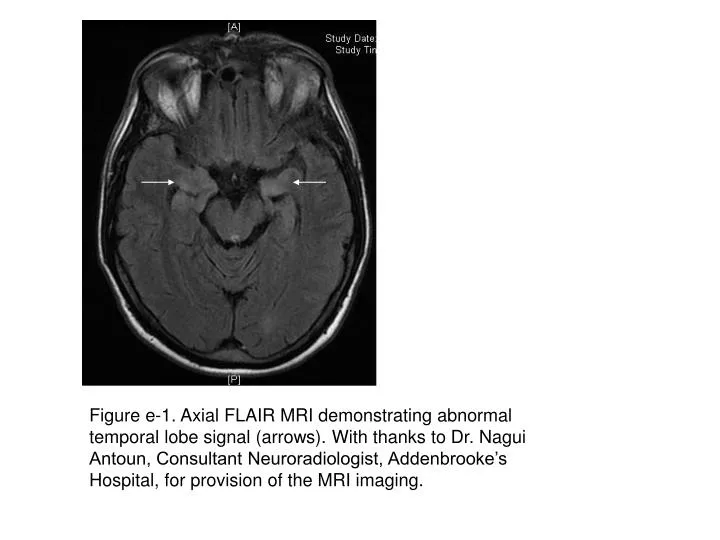
Figure e-1. Axial FLAIR MRI demonstrating abnormal temporal lobe signal (arrows). With thanks to Dr. Nagui Antoun, Consultant Neuroradiologist, Addenbrooke’s Hospital, for provision of the MRI imaging.